
Journal of Shanghai Jiao Tong University (Medical Science) ›› 2026, Vol. 46 ›› Issue (9): 1252-1259.doi: 10.3969/j.issn.1674-8115.2026.09.010
• Clinical research • Previous Articles
Xiao Hua, Zhang Zimin, Jiang Mengda, Guo Jiuhong( )
)
Received:2026-03-05
Accepted:2026-06-01
Online:2026-09-28
Published:2026-09-28
Contact:
Guo Jiuhong
E-mail:122708336@qq.com
CLC Number:
Xiao Hua, Zhang Zimin, Jiang Mengda, Guo Jiuhong. Multimodal functional magnetic resonance imaging combined with Ki-67 index for assessing pathological typing of head and neck rhabdomyosarcoma[J]. Journal of Shanghai Jiao Tong University (Medical Science), 2026,(9): 1252-1259.
Add to citation manager EndNote|Ris|BibTeX
URL: https://xuebao.shsmu.edu.cn/EN/10.3969/j.issn.1674-8115.2026.09.010
| Pathological type | E-RMS (n=21) | A-RMS (n=14) | SS-RMS (n=15) |
|---|---|---|---|
| Age/year | 13.3±8.7 | 21.8±12.8 | 17.4±15.2 |
| Sex/n(%) | |||
| Female | 9 (42.9) | 4 (28.6) | 2 (13.3) |
| Male | 12 (57.1) | 10 (71.4) | 13 (86.7) |
| Location/n(%) | |||
| Masticator space | 7 (33.3) | 1 (7.1) | 8 (53.3) |
| Orbit | 9 (42.9) | 2 (14.3) | 2 (13.3) |
| Nasal cavity & paranasal sinuses | 0 (0) | 8 (57.1) | 0 (0) |
| Nasal region | 0 (0) | 0 (0) | 3 (20.0) |
| Masseter muscle | 0 (0) | 1 (7.1) | 1 (6.7) |
| Buccal region | 2 (9.5) | 0 (0) | 0 (0) |
| Lip | 0 (0) | 2 (14.3) | 0 (0) |
| Parotid gland | 1 (4.8) | 0 (0) | 0 (0) |
| Mandible | 0 (0) | 0 (0) | 1 (6.7) |
| Palate | 1 (4.8) | 0 (0) | 0 (0) |
| Temporal bone | 1 (4.8) | 0 (0) | 0 (0) |
Tab 1 Demographic and clinical characteristics of RMS patients with different pathological types
| Pathological type | E-RMS (n=21) | A-RMS (n=14) | SS-RMS (n=15) |
|---|---|---|---|
| Age/year | 13.3±8.7 | 21.8±12.8 | 17.4±15.2 |
| Sex/n(%) | |||
| Female | 9 (42.9) | 4 (28.6) | 2 (13.3) |
| Male | 12 (57.1) | 10 (71.4) | 13 (86.7) |
| Location/n(%) | |||
| Masticator space | 7 (33.3) | 1 (7.1) | 8 (53.3) |
| Orbit | 9 (42.9) | 2 (14.3) | 2 (13.3) |
| Nasal cavity & paranasal sinuses | 0 (0) | 8 (57.1) | 0 (0) |
| Nasal region | 0 (0) | 0 (0) | 3 (20.0) |
| Masseter muscle | 0 (0) | 1 (7.1) | 1 (6.7) |
| Buccal region | 2 (9.5) | 0 (0) | 0 (0) |
| Lip | 0 (0) | 2 (14.3) | 0 (0) |
| Parotid gland | 1 (4.8) | 0 (0) | 0 (0) |
| Mandible | 0 (0) | 0 (0) | 1 (6.7) |
| Palate | 1 (4.8) | 0 (0) | 0 (0) |
| Temporal bone | 1 (4.8) | 0 (0) | 0 (0) |
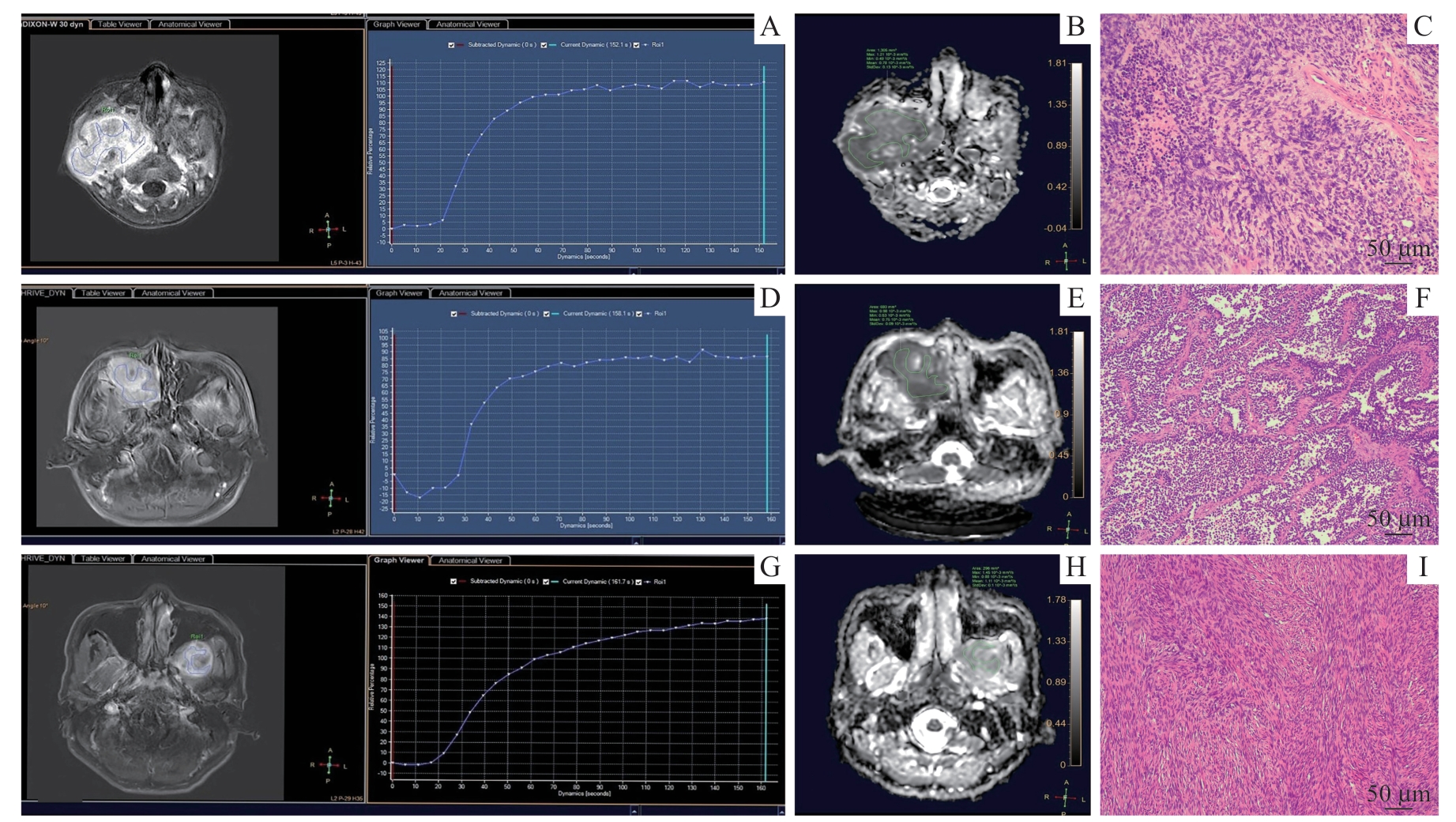
Fig 1 Functional MRI features of head and neck rhabdomyosarcoma and the corresponding pathological images
| Parameter | ICC (95%CI)/κ (95%CI) | |
|---|---|---|
| Inter-reader | Intra-reader | |
| ADC value | 0.918 (0.866‒0.951) | 0.902 (0.847‒0.941) |
| TIC type | 0.876 (0.812‒0.940) | 0.935 (0.891‒0.964) |
Tab 2 Inter- and intra-reader agreement of multimodal functional MRI parameters in RMS
| Parameter | ICC (95%CI)/κ (95%CI) | |
|---|---|---|
| Inter-reader | Intra-reader | |
| ADC value | 0.918 (0.866‒0.951) | 0.902 (0.847‒0.941) |
| TIC type | 0.876 (0.812‒0.940) | 0.935 (0.891‒0.964) |
| Parameter | E-RMS (n=21) | A-RMS (n=14) | SS-RMS (n=15) | H/χ2 value | P value |
|---|---|---|---|---|---|
| ADC value/(×10-3 mm2·s-1) | 0.890 (0.770, 1.060) | 0.745 (0.658, 0.845) | 1.010 (1.010, 1.300) | 26.688 | <0.001 |
| TIC type/n(%) | 6.845 | 0.031 | |||
| Ⅰ | 5 (23.8) | 5 (35.7) | 10 (66.7) | ||
| Ⅱ | 16 (76.2) | 9 (64.3) | 5 (33.3) | ||
| Ki-67 index/% | 40.000 (30.000, 60.000) | 40.000 (37.500, 50.000) | 20.000 (15.000, 30.000) | 17.019 | <0.001 |
Tab 3 Comparison of multimodal functional MRI parameters and Ki-67 index among three pathological types of RMS
| Parameter | E-RMS (n=21) | A-RMS (n=14) | SS-RMS (n=15) | H/χ2 value | P value |
|---|---|---|---|---|---|
| ADC value/(×10-3 mm2·s-1) | 0.890 (0.770, 1.060) | 0.745 (0.658, 0.845) | 1.010 (1.010, 1.300) | 26.688 | <0.001 |
| TIC type/n(%) | 6.845 | 0.031 | |||
| Ⅰ | 5 (23.8) | 5 (35.7) | 10 (66.7) | ||
| Ⅱ | 16 (76.2) | 9 (64.3) | 5 (33.3) | ||
| Ki-67 index/% | 40.000 (30.000, 60.000) | 40.000 (37.500, 50.000) | 20.000 (15.000, 30.000) | 17.019 | <0.001 |
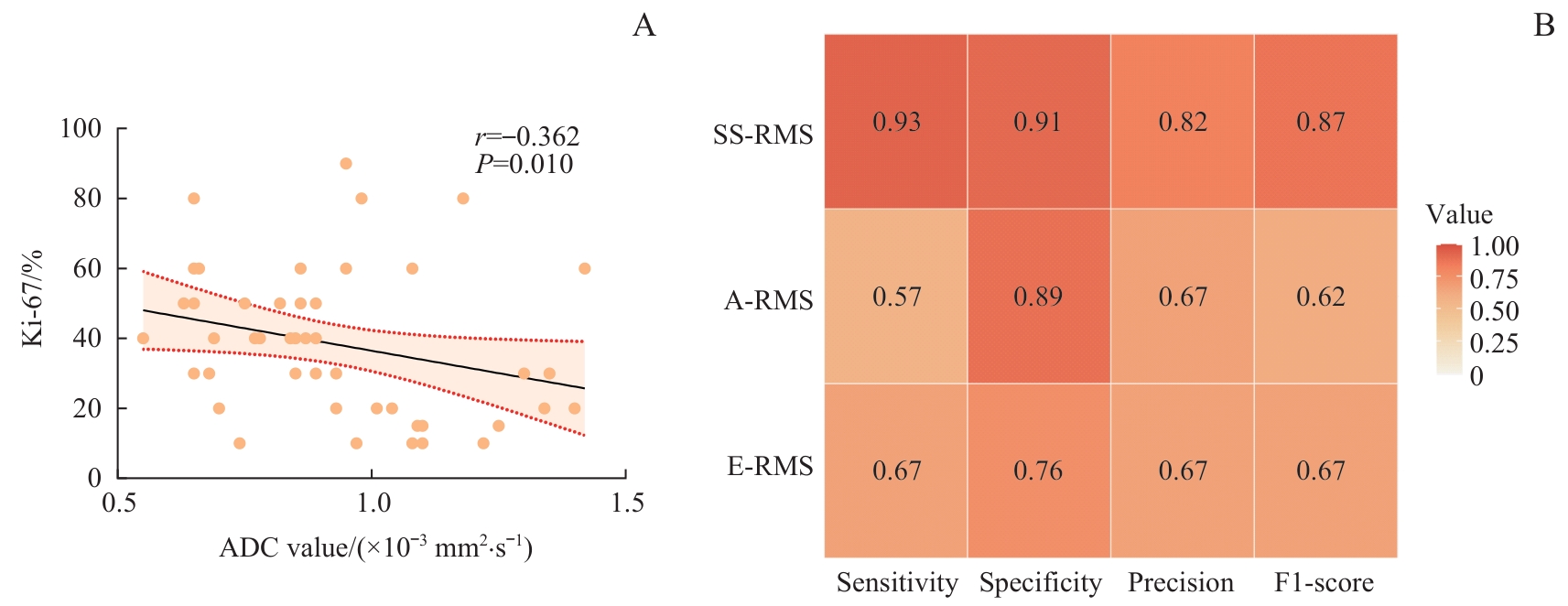
Fig 2 Correlation between ADC values and Ki-67 index (A), and performance matrix of the model combining multimodal functional MRI parameters with Ki-67 index for predicting different pathological types of RMS (B)
| Parameter | B | OR (95%CI) | P value |
|---|---|---|---|
| A-RMS① | |||
| ADC value/(×10-3 mm2·s-1) | -8.841 | 0 (0‒0.154) | 0.013 |
| TIC type | 0.164 | 1.178 (0.216‒6.441) | 0.850 |
| Ki-67 index/% | -0.026 | 0.975 (0.925‒1.027) | 0.331 |
| SS-RMS① | |||
| ADC value/(×10-3 mm2·s-1) | 7.634 | 2 067.165 (6.461‒661 414.539) | 0.009 |
| TIC type | 0.886 | 2.425 (0.242‒24.344) | 0.452 |
| Ki-67 index/% | -0.079 | 0.924 (0.862‒0.991) | 0.027 |
Tab 4 Multinomial Logistic regression analysis of RMS pathological subtypes based on multimodal functional MRI parameters combined with Ki-67 index
| Parameter | B | OR (95%CI) | P value |
|---|---|---|---|
| A-RMS① | |||
| ADC value/(×10-3 mm2·s-1) | -8.841 | 0 (0‒0.154) | 0.013 |
| TIC type | 0.164 | 1.178 (0.216‒6.441) | 0.850 |
| Ki-67 index/% | -0.026 | 0.975 (0.925‒1.027) | 0.331 |
| SS-RMS① | |||
| ADC value/(×10-3 mm2·s-1) | 7.634 | 2 067.165 (6.461‒661 414.539) | 0.009 |
| TIC type | 0.886 | 2.425 (0.242‒24.344) | 0.452 |
| Ki-67 index/% | -0.079 | 0.924 (0.862‒0.991) | 0.027 |
| Ground truth | Predicted value | Accuracy/% | ||||
|---|---|---|---|---|---|---|
| E-RMS | A-RMS | SS-RMS | ||||
| Total | ‒ | ‒ | ‒ | 72.0 | ||
| E-RMS | 14 | 4 | 3 | 66.7 | ||
| A-RMS | 6 | 8 | 0 | 57.1 | ||
| SS-RMS | 1 | 0 | 14 | 93.3 | ||
Tab 5 Accuracy of the model combining multimodal functional MRI parameters with Ki-67 index in predicting different pathological types of RMS
| Ground truth | Predicted value | Accuracy/% | ||||
|---|---|---|---|---|---|---|
| E-RMS | A-RMS | SS-RMS | ||||
| Total | ‒ | ‒ | ‒ | 72.0 | ||
| E-RMS | 14 | 4 | 3 | 66.7 | ||
| A-RMS | 6 | 8 | 0 | 57.1 | ||
| SS-RMS | 1 | 0 | 14 | 93.3 | ||
| [1] | 中国抗癌协会小儿肿瘤专业委员会, 中华医学会儿科学分会血液学组, 中华医学会小儿外科学分会肿瘤组. 中国儿童及青少年横纹肌肉瘤诊疗建议(CCCG-RMS-2016)[J]. 中华儿科杂志, 2017, 55(10): 724-728. |
| Chinese Children′s Cancer Group, Chinese Anti-Cancer Association, the Subspecialty Group of Hematology, the Society of Pediatrics, Chinese Medical Association, the Subspecialty Group of Oncology, the Society of Pediatric Surgery, Chinese Medical Association. Recommendations for the diagnosis and treatment of rhabdomyosarcoma in Chinese children and adolescents (CCCG-RMS-2016)[J]. Chinese Journal of Pediatrics, 2017, 55(10): 724-728. | |
| [2] | Darwish C, Shim T, Sparks A D, et al. Pediatric head and neck rhabdomyosarcoma: an analysis of treatment and survival in the United States (1975‒2016)[J]. Int J Pediatr Otorhinolaryngol, 2020, 139: 110403. |
| [3] | Sbaraglia M, Bellan E, Dei Tos A P. The 2020 WHO classification of soft tissue tumours: news and perspectives[J]. Pathologica, 2021, 113(2): 70-84. |
| [4] | 中华医学会病理学分会儿科病理学组, 中国抗癌协会小儿肿瘤专业委员会病理学组, 福棠儿童医学发展研究中心病理专业委员会. 儿童横纹肌肉瘤病理诊断规范化专家共识[J]. 中华病理学杂志, 2021, 50(10): 1110-1115. |
| Expert Group of Pediatrics of Chinese Society of Pathology, Expert Group of Pathology of Chinese Children Cancer Group, Expert Group of Pathology Committee of Futang Research Center of Pediatric Development. Consensus on the diagnosis of pediatric rhabdomyosarcoma[J]. Chinese Journal of Pathology, 2021, 50(10): 1110-1115. | |
| [5] | Gennaro N, Marrari A, Renne S L, et al. Multimodality imaging of adult rhabdomyosarcoma: the added value of hybrid imaging[J]. Br J Radiol, 2020, 93(1112): 20200250. |
| [6] | Agaram N P. Evolving classification of rhabdomyosarcoma[J]. Histopathology, 2022, 80(1): 98-108. |
| [7] | Wang X Y, Song L Y, Chong V, et al. Multiparametric MRI findings of sinonasal rhabdomyosarcoma in adults with comparison to carcinoma[J]. J Magn Reson Imaging, 2017, 45(4): 998-1004. |
| [8] | Inarejos Clemente E J, Navallas M, Barber Martínez de la Torre I, et al. MRI of rhabdomyosarcoma and other soft-tissue sarcomas in children[J]. Radiographics, 2020, 40(3): 791-814. |
| [9] | Parlak Ş, Gümeler E, Bulut E. Pediatric head and neck rhabdomyosarcoma; the role of MRI in differentiation from other common pediatric malignant head and neck tumors[J]. Turk J Pediatr, 2022, 64(3): 519-530. |
| [10] | Saboo S S, Krajewski K M, Zukotynski K, et al. Imaging features of primary and secondary adult rhabdomyosarcoma[J]. Am J Roentgenol, 2012, 199(6): W694-W703. |
| [11] | Chhabra A, Ashikyan O, Slepicka C, et al. Conventional MR and diffusion-weighted imaging of musculoskeletal soft tissue malignancy: correlation with histologic grading[J]. Eur Radiol, 2019, 29(8): 4485-4494. |
| [12] | Yuan J H, Xie D S, Fang S B, et al. Qualitative and quantitative MRI analysis of alveolar soft part sarcoma: correlation with histological grade and Ki-67 expression[J]. Insights Imaging, 2024, 15(1): 142. |
| [13] | Yuan Y, Zeng D W, Liu Y J, et al. DWI and IVIM are predictors of Ki67 proliferation index: direct comparison of MRI images and pathological slices in a murine model of rhabdomyosarcoma[J]. Eur Radiol, 2020, 30(3): 1334-1341. |
| [14] | Shomal Zadeh F, Pooyan A, Alipour E, et al. Dynamic contrast-enhanced magnetic resonance imaging (DCE-MRI) in differentiation of soft tissue sarcoma from benign lesions: a systematic review of literature[J]. Skeletal Radiol, 2024, 53(7): 1343-1357. |
| [15] | Vallières M, Serban M, Benzyane I, et al. Investigating the role of functional imaging in the management of soft-tissue sarcomas of the extremities[J]. Phys Imaging Radiat Oncol, 2018, 6: 53-60. |
| [16] | Kataoka M, Honda M, Sagawa H, et al. Ultrafast dynamic contrast-enhanced MRI of the breast: from theory to practice[J]. J Magn Reson Imaging, 2024, 60(2): 401-416. |
| [17] | Del Grande F, Subhawong T, Weber K, et al. Detection of soft-tissue sarcoma recurrence: added value of functional MR imaging techniques at 3.0 T[J]. Radiology, 2014, 271(2): 499-511. |
| [18] | Lv C L, Xue X L, Huang M G, et al. The dynamic contrast enhanced-magnetic resonance imaging and diffusion-weighted imaging features of alveolar soft part sarcoma[J]. Quant Imaging Med Surg, 2023, 13(10): 7269-7280. |
| [19] | Lee J H, Yoon Y C, Seo S W, et al. Soft tissue sarcoma: DWI and DCE-MRI parameters correlate with Ki-67 labeling index[J]. Eur Radiol, 2020, 30(2): 914-924. |
| [20] | Yang B T, Wang Y Z, Dong J Y, et al. MRI study of solitary fibrous tumor in the orbit[J]. Am J Roentgenol, 2012, 199(4): W506-W511. |
| [21] | Soffer S, Amitai M M, Shimon O, et al. Rhabdomyosarcoma disease spread evaluation on CT scans: association with primary tumor size and Ki-67 proliferation marker[J]. Clin Imag, 2019, 56: 41-46. |
| [22] | McCarville M B. What MRI can tell us about neurogenic tumors and rhabdomyosarcoma[J]. Pediatr Radiol, 2016, 46(6): 881-890. |
| [23] | van Ewijk R, Chatziantoniou C, Adams M, et al. Quantitative diffusion-weighted MRI response assessment in rhabdomyosarcoma: an international retrospective study on behalf of the European paediatric Soft tissue sarcoma Study Group Imaging Committee[J]. Pediatr Radiol, 2023, 53(12): 2539-2551. |
| [24] | Liu J H, Qi M, Huang W H, et al. The magnetic resonance characteristics of sinonasal rhabdomyosarcoma in adults: analysis of 27 cases and comparison with pathological subtypes[J]. BMC Med Imaging, 2023, 23(1): 98. |
| [25] | Zeng J J, Liu L, Li J Y, et al. MRI features of different types of sinonasan rhabdomyosarcomas: a series of eleven cases[J]. Dentomaxillofac Radiol, 2021, 50(8): 20210030. |
| [26] | Yao R, Cheng A L, Zhang Z W, et al. Correlation between apparent diffusion coefficient and the Ki-67 proliferation index in grading pediatric glioma[J]. J Comput Assist Tomogr, 2023, 47(2): 322-328. |
| [27] | Sun M T, Cheng J L, Zhang Y, et al. Application of DWIBS in malignant lymphoma: correlation between ADC values and Ki-67 index[J]. Eur Radiol, 2018, 28(4): 1701-1708. |
| [1] | Yin Wenyu, Wang Ruijie, Ma Hailong, Jin Shufang. Study on how histone lysine lactylation regulates zinc finger protein 668 to facilitate proliferation of head and neck squamous cell carcinoma cells [J]. Journal of Shanghai Jiao Tong University (Medical Science), 2026, 46(2): 151-162. |
| [2] | LI Siyu, CHEN Ya, HU Wentao, DAI Yongming, WU Yingwei. Using diffusion-relaxation correlation spectroscopic imaging to assess the heterogeneity of head and neck tumors and identify occult lymph node metastasis [J]. Journal of Shanghai Jiao Tong University (Medical Science), 2025, 45(9): 1202-1213. |
| [3] | CHEN Yinan, ZHENG Yang, ZENG Hanlin, LEI Ming. Mechanism of Fas-associated protein with death domain in promoting proliferation of head and neck squamous cell carcinoma cells [J]. Journal of Shanghai Jiao Tong University (Medical Science), 2025, 45(4): 404-414. |
| [4] | AN Junyi, CHEN Biying, CHEN Xunrui, YIN Shanshan, BIAN Zhouliang, LIU Feng. SFXN3 expression in head and neck squamous cell carcinoma and its effect on cell proliferation [J]. Journal of Shanghai Jiao Tong University (Medical Science), 2024, 44(4): 427-434. |
| [5] | GU Fen, WANG Yueping, YANG Wenyu, ZHAO Xiaomei, TANG Yan, SHEN Shukun, MAO Yan, ZHANG Jinfeng, WU Yifan, ZHANG Yuanyuan, YANG Yue, ZHANG Jianchun, YU Hong, WANG Lan, HAO Guihua, HOU Lili. Expert consensus on postoperative rehabilitation nursing of patients with head and neck cancer [J]. Journal of Shanghai Jiao Tong University (Medical Science), 2023, 43(10): 1289-1296. |
| [6] | CHEN Weihong, HOU Lili, YANG Ling, MAO Yan, ZHANG Jinfeng. Efficacy of preventive intervention of cryotherapy on radiation-induced oral mucositis in head and neck cancer: a meta-analysis [J]. Journal of Shanghai Jiao Tong University (Medical Science), 2022, 42(5): 635-645. |
| [7] | Yu-sheng LU, Wen-yi YANG, Hai-long MA, Jing-zhou HU. Impact of Ras association domain family 5 on cell migration and invasion of head and neck squamous cell carcinoma [J]. JOURNAL OF SHANGHAI JIAOTONG UNIVERSITY (MEDICAL SCIENCE), 2021, 41(6): 717-723. |
| [8] | Hao GU, Xi YANG, Zi-min ZHANG, Yun-bo JIN, Li HU, Hui CHEN, Xiao-xi LIN. Image-fusion-based surgical navigation in sclerotherapy for low-flow vascular malformations in the risky regions of head and neck [J]. JOURNAL OF SHANGHAI JIAOTONG UNIVERSITY (MEDICAL SCIENCE), 2021, 41(5): 695-700. |
| [9] | Tong TONG, Xing QIN, Fei XIE, Ying-ying JIANG, Jian-bo SHI, Jian-jun ZHANG. USP47 induces cisplatin resistance in head and neck squamous cell carcinoma by up-regulation of anti-apoptotic proteins [J]. JOURNAL OF SHANGHAI JIAOTONG UNIVERSITY (MEDICAL SCIENCE), 2021, 41(4): 434-441. |
| [10] | ZHANG Yu-han, CHEN Xue-hua, ZHANG Hao. Down-regulation of aurora kinase A activity inhibits pro-angiogenesis, migration and invasion of Hep2 cells [J]. , 2018, 38(10): 1181-. |
| [11] | LIU Kai, TAN Yao, LI Ya-wei, et al. Analysis of gene expression and polymorphism of allele HLA-A*02 of Han patients with nasopharyngeal carcinoma in Xinjiang [J]. , 2015, 35(1): 59-. |
| [12] | LIU Kai, MA Ling-ling, WANG Duo-ming, et al. Relationship between HPV infection and expression of HIF-1alpha and VEGF in head and neck squamous cell carcinoma and its prognostic value [J]. , 2013, 33(6): 806-. |
| Viewed | ||||||
|
Full text |
|
|||||
|
Abstract |
|
|||||
