上海交通大学学报(医学版), 2025, 45(6): 727-734 doi: 10.3969/j.issn.1674-8115.2025.06.007

论著 · 基础研究

大黄素改善阿尔茨海默病认知障碍、内质网应激和神经炎症的研究

杨乐1, 周怡1, 王钶韵2, 赖娅莉,1

1.成都医学院第二附属医院,核工业四一六医院神经内科,成都 610041

2.四川省医学科学院·四川省人民医院神经内科,成都 610072

Research on the improvement of cognitive impairment, endoplasmic reticulum stress and neuroinflammation in Alzheimer's disease by emodin

YANG Le1, ZHOU Yi1, WANG Keyun2, LAI Yali,1

1.Department of Neurology, The Second Affiliated Hospital of Chengdu Medical College, 416 Nuclear Industry Hospital, Chengdu 610041, China

2.Department of Neurology, Sichuan Academy of Medical Sciences and Sichuan Provincial People's Hospital, Chengdu 610072, China

通讯作者: 赖娅莉,副主任医师,硕士;电子信箱:838856089@qq.com

编委: 徐敏

收稿日期: 2024-11-18   接受日期: 2025-02-18   网络出版日期: 2025-06-28

基金资助: 2020年四川省卫生健康科研课题立项项目.  20ZD009

Corresponding authors: LAI Yali, E-mail:838856089@qq.com.

Received: 2024-11-18   Accepted: 2025-02-18   Online: 2025-06-28

作者简介 About authors

杨乐(1988—),男,副主任医师,硕士;电子信箱:yulele594@163.com。 。

摘要

目的·探讨大黄素在阿尔茨海默病(Alzheimer's disease,AD)中的作用及其潜在机制。方法·将野生型C57BL/6J小鼠和3×Tg-AD小鼠分为6组:对照组(C57BL/6J小鼠)、AD组(3×Tg-AD小鼠)、大黄素25 mg/kg组(3×Tg-AD小鼠+大黄素25 mg/kg)、大黄素50 mg/kg组(3×Tg-AD小鼠+大黄素50 mg/kg)、大黄素100 mg/kg组(3×Tg-AD小鼠+大黄素100 mg/kg)和多奈哌齐组(3×Tg-AD小鼠+多奈哌齐3 mg/kg)。采用Morris水迷宫实验分析小鼠学习记忆能力,采用免疫组织化学法检测胶质纤维酸性蛋白(glial fibrillary acidic protein,GFAP)、葡萄糖调节蛋白78(glucose-regulated protein 78kDa,GRP78)和肌醇需求酶1α(inositol-requiring enzyme 1α,IRE1α)的表达,采用酶联免疫吸附试验(enzyme-linked immunosorbent assay,ELISA)分析脑组织中肿瘤坏死因子-α(tumor necrosis factor-α,TNF-α)、白介素-1β(interleukin-1β,IL-1β)和IL-6的含量,采用Western blotting检测NF-κB p65、p-NF-κB p65、p38、p-p38的蛋白表达。结果·与对照组相比,AD组小鼠认知能力下降,GFAP表达升高,TNF-α、IL-1β、IL-6含量增多,GRP78和IRE1α表达升高,NF-κB p65、p38磷酸化表达升高。与AD组相比,大黄素改善AD小鼠的认知能力障碍,抑制星形胶质细胞过度激活和神经炎症,并降低脑组织GRP78、IRE1α、磷酸化NF-κB p65和磷酸化p38的表达。结论·大黄素能有效改善AD小鼠认知能力障碍,其机制可能与抑制星形胶质细胞内质网应激介导的神经炎症有关。

关键词: 阿尔茨海默病 ; 大黄素 ; 神经炎症 ; 内质网应激

Abstract

Objective ·To explore the effects and potential mechanisms of emodin on Alzheimer's disease (AD). Methods ·Wild-type C57BL/6J mice and 3×Tg-AD mice were divided into 6 groups: Control group (C57BL/6J mice), AD group (3×Tg-AD mice), Emodin 25 mg/kg group (3×Tg-AD mice + Emodin 25 mg/kg), Emodin 50 mg/kg group (3×Tg-AD mice + Emodin 50 mg/kg), Emodin 100 mg/kg group (3×Tg-AD mice + Emodin 100 mg/kg) and Donepezil group (3×Tg-AD mice + Donepezil 3 mg/kg). The Morris water maze test was used to evaluate the learning and memory abilities of mice. The expression of glial fibrillary acidic protein (GFAP), glucose-regulated protein 78kDa (GRP78), and inositol-requiring enzyme 1α (IRE1α) was detected by immunohistochemistry. The levels of tumor necrosis factor-α (TNF-α), interleukin-1β (IL-1β), and IL-6 in brain tissue were measured by enzyme-linked immunosorbent assay (ELISA). Western blotting was used to detect the expression of NF-κB p65, p-NF-κB p65, p38, and p-p38 proteins. Results ·Compared with the control group, mice in the AD group showed impaired cognition, increased GFAP expression, elevated levels of TNF-α, IL-1β and IL-6, and increased expression of GRP78 and IRE1α, along with enhanced phosphorylation of NF-κB p65 and p38. Compared with the AD group, emodin improved cognitive impairment of AD mice, inhibited astrocyte overactivation and neuroinflammation, and decreased the expression of GRP78, IRE1α, phosphorylated NF-κB p65, and phosphorylated p38 in brain tissue. Conclusion ·Emodin can effectively improve cognitive impairment in AD mice, which may be related to the inhibition of endoplasmic reticulum stress-mediated neuroinflammation in astrocytes.

Keywords: Alzheimer's disease (AD) ; emodin ; neuroinflammation ; endoplasmic reticulum stress

PDF (4800KB) 元数据 多维度评价 相关文章 导出 EndNote| Ris| Bibtex  收藏本文

本文引用格式

杨乐, 周怡, 王钶韵, 赖娅莉. 大黄素改善阿尔茨海默病认知障碍、内质网应激和神经炎症的研究. 上海交通大学学报(医学版)[J], 2025, 45(6): 727-734 doi:10.3969/j.issn.1674-8115.2025.06.007

YANG Le, ZHOU Yi, WANG Keyun, LAI Yali. Research on the improvement of cognitive impairment, endoplasmic reticulum stress and neuroinflammation in Alzheimer's disease by emodin. Journal of Shanghai Jiao Tong University (Medical Science)[J], 2025, 45(6): 727-734 doi:10.3969/j.issn.1674-8115.2025.06.007

阿尔茨海默病(Alzheimer's disease,AD)是一种认知记忆丧失的神经退行性疾病,主要发生在老年人群1-2。全世界有超过5 000万患者受到AD的影响,预计到2050年这一数字将增加1倍3。AD的表型特征是记忆力丧失、社会行为受损和人格改变24。AD的病因至今尚未清晰,治疗效果也不明显。因此,探索AD的新治疗策略具有极大的必要性和临床价值。大黄素(emodin,CAS号:518-82-1)是从中药大黄(蓼科多年生草本植物掌叶大黄的根及根茎)中提取的一种天然化合物,具有抗炎、抗氧化和抗肿瘤等特性5-8。研究9发现,大黄素可显著抑制AD小鼠海马区氧化应激和神经炎症。持续性内质网(endoplasmic reticulum,ER)应激被认为是许多慢性疾病的驱动因素。在神经退行性疾病中,由ER应激引起的反应可导致神经元功能障碍。细胞对ER应激的反应是通过诱导未折叠蛋白反应(unfolded protein response,UPR)来启动,且在大量的AD患者的大脑样本中观察到UPR的上调10-11。然而,大黄素是否影响AD的ER应激仍有待进一步阐明。鉴于此,本研究假设大黄素在AD中的保护作用是通过抑制星形胶质细胞ER应激介导的神经炎症而实现,拟通过动物模型对其进行验证。

1 对象与方法

1.1 药物与试剂

大黄素(纯度≥90.0%)购自美国Sigma公司,多奈哌齐购自美国MCE生物公司。肿瘤坏死因子-α(tumor necrosis factor-α,TNF-α)、白介素-1β(interleukin-1β,IL-1β)、IL-6、酶联免疫吸附实验(enzyme-linked immunosorbent assay,ELISA)试剂盒购自武汉云克隆生物公司,免疫荧光双染试剂购自北京索莱宝科技有限公司,4',6-二脒基-2-苯基吲哚(4',6-diamidino-2-phenylindole,DAPI)购自美国Thermo Fisher生物公司,胶质纤维酸性蛋白(glial fibrillary acidic protein,GFAP)抗体、葡萄糖调节蛋白78(glucose-regulated protein 78kDa,GRP78)抗体、肌醇需求酶1α(inositol-requiring enzyme 1α,IRE1α)抗体、NF-κB p65抗体、p-NF-κB p65抗体、p38抗体和p-p38抗体购自英国 Abcam公司。

1.2 实验动物

8月龄3×Tg-AD基因纯合子小鼠购自常州卡文斯实验动物有限公司,使用许可证号:SCXK(苏)2021-0013。8月龄野生型C57BL/6J小鼠购自上海生物医药技术研究院,使用许可证号:SYXK(沪)2024-0017。所有动物被饲养在12 h光照/暗循环的环境中,可以随意获得食物和水。

1.3 实验设计

将野生型C57BL/6J小鼠和3×Tg-AD小鼠分为6组(n=6):对照组(C57BL/6J小鼠)、AD组(3×Tg-AD小鼠)、大黄素25 mg/kg组(3×Tg-AD小鼠+大黄素25 mg/kg)、大黄素50 mg/kg组(3×Tg-AD小鼠+大黄素50 mg/kg)、大黄素100 mg/kg组(3×Tg-AD小鼠+大黄素100 mg/kg)、多奈哌齐组(3×Tg-AD小鼠+多奈哌齐3 mg/kg)。大黄素和多奈哌齐的剂量参考之前的报道912。大黄素各剂量均溶解于0.5% 羧甲基纤维素钠(CMC-Na)中,小鼠每日经腹腔分别注射1次(25、50、100 mg/kg),持续4周。多奈哌齐组,将多奈哌齐溶解于0.5% CMC-Na中,每日口服给药(3 mg/kg),持续4周。对照组和AD组腹腔注射同体积的无菌0.9%生理盐水。最后一次大黄素干预后,进行5 d的行为学测试,之后处死小鼠,收集新鲜脑组织,部分用于收集匀浆上清液和分离蛋白,剩余部分用于制备石蜡切片。

1.4 Morris水迷宫实验

将小鼠置于圆形水池中的不同起始位置,水温(25±1)℃。在第二象限设置一个平台,水面以下距离为1 cm。行为学测试每日进行2次,连续5 d。将小鼠面向池壁置于水中,并在90 s内探索隐藏平台。当小鼠成功找到平台并在平台上停留2 s时,记录其逃跑潜伏期。如果小鼠在90 s内没有找到平台,则记录潜伏期为90 s,引导小鼠到平台并在平台上停留15 s。在最后1次试验训练后24 h进行探针试验,取下平台,将小鼠面朝池壁置于水中,让小鼠自由寻找平台90 s,记录穿越平台位置的次数。

1.5 免疫组织化学实验

取储存的脑组织制备石蜡切片,经脱蜡、再水化,使用加热的柠檬酸盐缓冲液(10 mmol/L,pH 6.0)修复抗原10 min。用3% H2O2孵育15 min以消除内源性过氧化物酶活性。随后在室温下用5%牛血清白蛋白封闭15 min。将切片与抗GFAP(1∶500)、抗GRP78(1∶600)和抗IRE1α(1∶600)一抗在4 ℃下孵育过夜。冲洗切片,在37 ℃下与HRP偶联的二抗共孵育1 h。经DAB显色处理,并用苏木精复染3 min。脱水、透明并封片,使用显微镜获取图像。

1.6 免疫荧光双染实验

将石蜡切片脱蜡,然后进行柠檬酸缓冲液抗原修复,并用5%牛血清白蛋白封闭15 min。将切片与抗GRP78和抗GFAP的一抗在4 ℃下孵育过夜。在室温下,与FITC/Cy3偶联二抗在暗室中孵育1 h。随后,用DAPI再处理切片20 min。冲洗并密封切片,使用荧光显微镜获取图像。

1.7 ELISA法检测脑组织炎症因子

使用匀浆器在PBS缓冲溶液中匀浆化脑组织,离心10 min并收集上清液。根据制造商说明,使用ELISA商业试剂盒分别检测TNF-α、IL-1β和IL-6在脑中的含量,用酶标仪在λ=450 nm处测定吸光度。

1.8 Western blotting检测NF-κB p65、p-NF-κB p65、p38、p-p38蛋白表达

取新鲜脑组织,提取蛋白质,并使用BCA试剂盒测定蛋白浓度。取20 μg蛋白样本,经SDS-PAGE电泳分离,转移至PVDF膜上。室温下,使用5%的脱脂牛奶封闭膜2 h,随后孵育抗NF-κB p65(1∶1 000)、抗p-NF-κB p65(1∶1 000)、抗p38(1∶2 000)、抗p-p38(1∶2 000)和抗β-actin(1∶500)一抗于4 ℃过夜。将膜与辣根过氧化物酶标记的对应二抗在室温下孵育1 h。使用化学发光成像技术进行曝光,并使用Image J软件分析蛋白条带,计算蛋白相对表达量。

1.9 统计学方法

采用SPSS 25.0软件分析数据。符合正态分布的定量资料以x±s表示。2组间的差异比较使用t检验,多组间的差异比较使用单因素方差分析。P<0.05表示差异具有统计学意义。

2 结果

2.1 大黄素改善AD小鼠认知能力

图1所示,与对照组相比,AD组小鼠游泳轨迹变简单、到达平台的潜伏期明显延长;与AD组相比,大黄素各剂量组和多奈哌齐组的小鼠游泳轨迹由简单变复杂、到达平台的潜伏期明显缩短。与对照组相比,AD组小鼠穿越平台次数、在目标象限内花费时间和总游泳距离明显减少;与AD组相比,大黄素各剂量组和多奈哌齐组穿越平台次数、在目标象限内花费时间和总游泳距离明显增加。

图1

图1   大黄素对AD小鼠认知能力障碍的影响

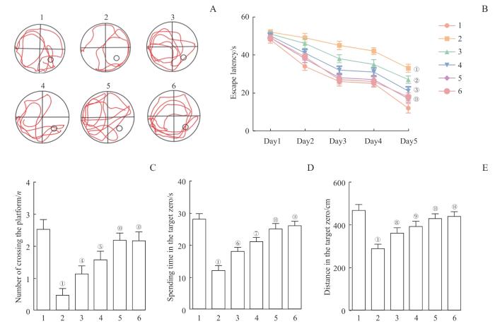
Note: A. Tracks of the mice in the probe trial. B. Escape latency in the Morris water maze test. C. Number of platform crossings. D. Time spent in the target quadrant. E. Total swimming distance in the target quadrant. 1—Control group, 2—AD group, 3—Emodin 25 mg/kg group, 4—Emodin 50 mg/kg group, 5—Emodin 100 mg/kg group, 6—Donepezil group. P<0.001, compared with the control group; P=0.015, P=0.003, P=0.022, P=0.001, P=0.038, P=0.004, P=0.033, P=0.007, P<0.001, compared with the AD group.

Fig 1   Effect of emodin on cognitive impairment in AD mice


2.2 大黄素抑制AD小鼠星形胶质细胞的过度激活

图2所示,与对照组相比,AD组小鼠大脑皮质中的GFAP阳性表达显著升高;与AD组相比,大黄素各剂量组和多奈哌齐组小鼠大脑皮质中的GFAP阳性表达显著降低。

图2

图2   大黄素对星形胶质细胞激活的影响

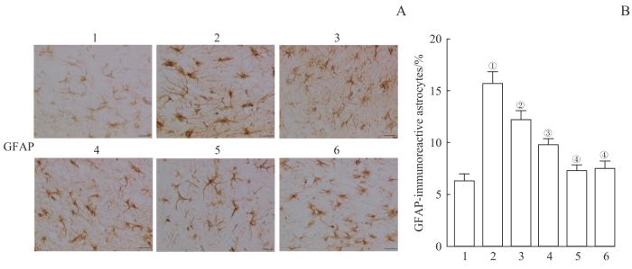
Note: A. Immunohistochemical detection of GFAP expression in the mouse cerebral cortex (×400). B. Percentage of GFAP-positive expression. 1—Control group, 2—AD group, 3—Emodin 25 mg/kg group, 4—Emodin 50 mg/kg group, 5—Emodin 100 mg/kg group, 6—Donepezil group. P<0.001, compared with the control group. P=0.016, P=0.002, P<0.001, compared with the AD group.

Fig 2   Effect of emodin on the activation of astrocytes


2.3 大黄素降低AD小鼠ER应激相关蛋白的表达

图3所示,与对照组相比,AD小鼠脑组织中的ER标记蛋白GRP78和IRE1α阳性表达显著增加;与AD组相比,大黄素各剂量组和多奈哌齐组小鼠脑组织中的GRP78和IRE1α阳性表达明显降低。与对照组相比,AD小鼠的脑组织中,星形胶质细胞标志物GFAP和ER标记物GRP78的表达增加并明显共定位;与AD组相比,大黄素各剂量组和多奈哌齐组明显降低了GFAP和GRP78的表达和共定位。

图3

图3   大黄素对ER应激相关蛋白表达的影响

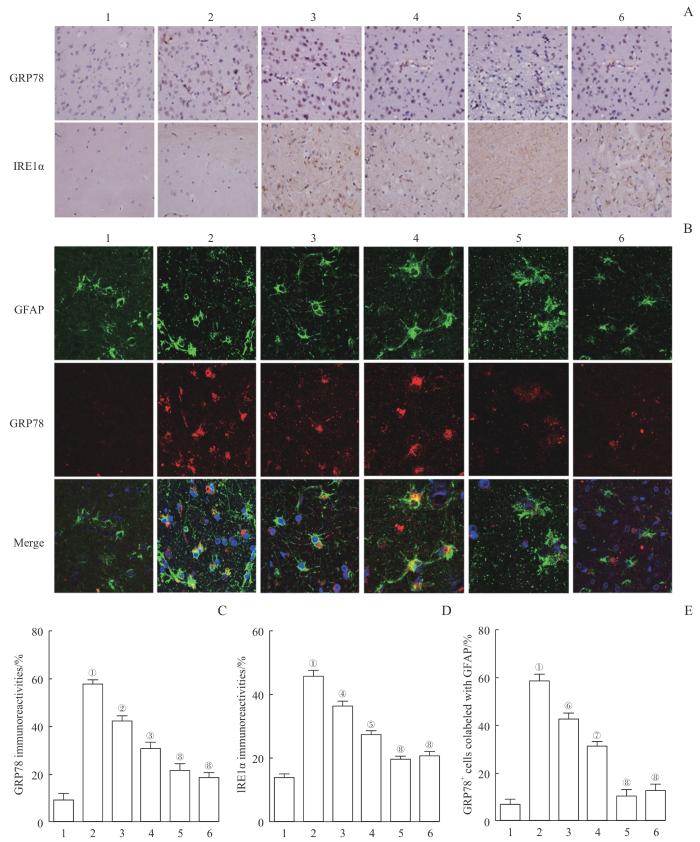
Note: A. Immunohistochemical staining of GRP78 and IRE1α in mouse cerebral cortex (×400). B. Double immunofluorescence staining of GFAP and GRP78 in mouse brain tissue (×400). C. Percentage of GRP78-positive expression. D. Percentage of IRE1α-positive expression. E. Percentage of GFAP-labeled GRP78+ cells.1—Control group, 2—AD group, 3—Emodin 25 mg/kg group, 4—Emodin 50 mg/kg group, 5—Emodin 100 mg/kg group, 6—Donepezil group. P<0.001, compared with the control group. P=0.027, P=0.005, P=0.018, P=0.002, P=0.013, P=0.009, P<0.001, compared with the AD group.

Fig 3   Effect of emodin on the expression of ER stress-related proteins


2.4 大黄素改善AD小鼠的炎症反应

图4所示,与对照组相比,AD小鼠组脑组织中TNF-α、IL-1β、IL-6含量显著增多。与AD组相比,大黄素各剂量组和多奈哌齐组脑组织中TNF-α、IL-1β、IL-6含量显著减少。

图4

图4   大黄素对AD小鼠炎症反应的影响

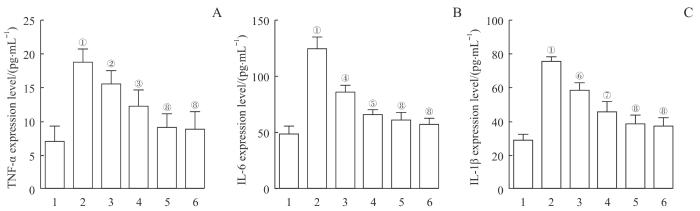
Note: A. TNF-α expression levels in brain tissue. B. IL-6 expression levels in brain tissue. C. IL-1β expression levels in brain tissue. 1—Control group, 2—AD group, 3—Emodin 25 mg/kg group, 4—Emodin 50 mg/kg group, 5—Emodin 100 mg/kg group, 6—Donepezil group. P<0.001, compared with the control group. P=0.031, P=0.004, P=0.037, P=0.008, P=0.043, P=0.003, P<0.001, compared with the AD group.

Fig 4   Effect of emodin on inflammatory response in AD mice


2.5 大黄素降低AD小鼠的炎症信号

通过Western blotting检测了各组2种经典的炎症信号,如图5所示。与对照组相比,AD组NF-κB p65、p38的磷酸化水平明显增加。与AD组相比,大黄素各剂量组和多奈哌齐组NF-κB p65、p38的磷酸化水平显著降低。

图5

图5   大黄素对炎症信号的影响

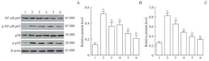
Note: A. Western blotting images of NF-κB p65, p-NF-κB p65, p38, and p-p38. B. Relative expression of p-NF-κB p65/NF-κB p65. C. Relative expression of p-p38/p38. 1—Control group, 2—AD group, 3—Emodin 25 mg/kg group, 4—Emodin 50 mg/kg group, 5—Emodin 100 mg/kg group, 6—Donepezil group. P<0.001, compared with the control group. P=0.035, P=0.042, P=0.028, P=0.007, P<0.001, compared with the AD group.

Fig 5   Effect of emodin on the inflammation signaling pathways


3 讨论

AD是一种复杂的、多因素诱导的进行性神经退行性疾病,以认知能力下降和行为改变为特征,是痴呆的最常见原因。虽然临床上使用他克林、利瓦司明、美金刚和多奈哌齐治疗AD,但它们只能暂时改善AD的症状13-14。因此,迫切需要开发安全有效的治疗AD的药物。本研究以3×Tg-AD小鼠作为AD模型,研究了大黄素对小鼠认知能力及神经炎症的影响。结果表明,大黄素显著改善了AD小鼠的认知能力和空间学习障碍,并有效抑制了AD小鼠的炎症反应,这与之前的研究结果一致9。尽管炎症被认为是身体的一种自然且有益的生理反应,它能促进碎片的清除和组织修复,但持续的炎症会损害神经元功能并导致细胞死亡15

星形胶质细胞作为中枢神经系统中占优势的常住细胞群,参与神经递质摄取和循环、炎症、离子平衡以及血脑屏障维持等,是神经元正常工作的重要微环境和维持大脑动态环境平衡的关键,因此成为干预AD等神经退行性疾病的研究热点16-17。AD的发病机制并不局限于中间神经元,也与脑内的神经胶质细胞密切相关18。星形胶质细胞病理反应以星形胶质细胞反应性增生为代表,表现为GFAP表达的显著增加19。本研究采用免疫组化方法检测小鼠大脑皮质星形胶质细胞中GFAP的表达。结果显示,AD小鼠大脑皮质中GFAP的表达明显高于对照组小鼠,表明该模型中反应性星形胶质细胞增加;而大黄素明显降低了AD小鼠的GFAP表达,提示大黄素可以抑制星形胶质细胞的过度激活。尽管已有研究20表明大黄素可以抑制LPS诱导的星形细胞瘤细胞的焦亡、炎症,但尚未见关于其对星形胶质细胞作用的报道。本研究局限于动物模型,需进一步结合细胞模型深入研究其活性机制。

在ER应激下,细胞通过激活UPR来逃避严重损伤21。AD患者脑样本中UPR-ER上调,提示ER应激在AD中的作用不容忽视22。UPR-ER包含3种应激反应跨膜蛋白的复杂网络,如RNA样ER激酶、IRE1和活化转录因子623。正常状态下,GRP78与这些跨膜蛋白结合以维持稳定的非反应状态。当损伤引起ER应激时,GRP78与ER分离并激活UPR应答24。因此,GRP78被认为是ER应激的重要标志物。在本研究中,免疫组化结果显示GRP78表达增加,表明AD模型中存在ER应激。此外,免疫荧光双染色结果显示,星形胶质细胞反应蛋白GFAP和GRP78在AD小鼠脑内明显共定位,提示小鼠大脑中的星形胶质细胞发生了ER应激。

在ER应激反应期间,蛋白激酶R样内质网激酶、IRE-1会触发炎症信号。其中,衔接蛋白TNF受体相关因子2 在IRE1α寡聚反应后被招募。复合物激活的炎症途径包括NF-κB和MAPK家族JNK和p38的蛋白质25-26。因此,本研究检测了IRE1α和p-NF-κB p65、NF-κB p65、p38、p-p38的蛋白表达。结果显示,AD小鼠中IRE1α、p-NF-κB p65和p-p38的蛋白表达显著升高,提示ER应激发生在AD小鼠中,并可能通过IRE1α-TRAF2轴介导NF-κB/p38炎症通路的激活,从而导致神经炎症。而大黄素治疗后,显著降低了IRE1α、p-NF-κB p65和p-p38的蛋白表达,表明大黄素对AD动物模型的ER应激激活和炎症信号通路具有明显的抑制作用。

综上,本研究表明大黄素通过抑制ER应激缓解AD小鼠的神经炎症,逆转小鼠的学习记忆障碍。研究结果为大黄素作为治疗AD的潜在候选药物提供了依据。尽管本研究有良好的数据,但外源性补充大黄素的抗AD潜力尚未具体确定。大黄素介导的抗AD作用的机制需要使用合适的模型系统进行更为深入的研究。此外,尚需开展临床试验,以评估大黄素对AD患者的益处;大黄素的毒性问题更需深入研究。

伦理批准和动物权利声明

本研究涉及的动物实验已取得四川省医学科学院·四川省人民医院伦理委员会批准(S2023-030-05)。实验操作过程严格遵守《实验动物护理和使用指南》。

Ethics Approval and Animal Right

The animal experiments involved in this study were approved by the Ethics Committee of Sichuan Academy of Medical Sciences and Sichuan Provincial People's Hospital (S2023-030-05). The experimental operation process strictly follows the Guidelines for the Care and Use of Experimental Animals.

作者贡献声明

杨乐、周怡参与实验设计,杨乐、周怡、王钶韵参与实验操作、数据采集及数据分析,杨乐、周怡、王钶韵、赖娅莉参与论文的写作和修改。所有作者均阅读并同意最终稿件的提交。

AUTHOR's CONTRIBUTIONS

YANG Le and ZHOU Yi participated in the experimental design. YANG Le, ZHOU Yi and WANG Keyun participated in experimental operations, data collection, and data analysis. YANG Le, ZHOU Yi, WANG Keyun and LAI Yali participated in the writing and editing of the paper. All authors have read and agreed to submit the final manuscript.

利益冲突声明

所有作者声明不存在利益冲突。

COMPETING INTERESTS

All authors disclose no relevant conflict of interests.

参考文献

JETT S, MALVIYA N, SCHELBAUM E, et al. Endogenous and exogenous estrogen exposures: how women's reproductive health can drive brain aging and inform Alzheimer's prevention[J]. Front Aging Neurosci, 2022, 14: 831807.

[本文引用: 1]

ZHAO M, WANG Y C, SHEN Y X, et al. A review of the roles of pathogens in Alzheimer's disease[J]. Front Neurosci, 2024, 18: 1439055.

[本文引用: 2]

AVILA J, PERRY G. A multilevel view of the development of Alzheimer's disease[J]. Neuroscience, 2021, 457: 283-293.

[本文引用: 1]

SONG L, PIAO Z Y, YAO L F, et al. Schisandrin ameliorates cognitive deficits, endoplasmic reticulum stress and neuroinflammation in streptozotocin (STZ)-induced Alzheimer's disease rats[J]. Exp Anim, 2020, 69(3): 363-373.

[本文引用: 1]

LIU Y H, SHANG L R, ZHOU J B, et al. Emodin attenuates LPS-induced acute lung injury by inhibiting NLRP3 inflammasome-dependent pyroptosis signaling pathway in vitro and in vivo[J]. Inflammation, 2022, 45(2): 753-767.

[本文引用: 1]

陈芬,袁飞飞,李伟,等.大黄素调节AMPK/TXNIP/NLRP3信号通路对子痫前期大鼠炎症反应的影响[J]. 中国临床药理学杂志, 2024, 40(14): 2068-2072.

CHEN F, YUAN F F, LI W, et al. Effects of emodin on inflammatory response in preeclampsia rats by regulating AMPK/TXNIP/NLRP3 signaling pathway[J]. Chinese Journal of Clinical Pharmacology, 2024, 40(14): 2068-2072.

XING M, MA X Y, WANG X, et al. Emodin disrupts the Notch1/Nrf2/GPX4 antioxidant system and promotes renal cell ferroptosis[J]. J Appl Toxicol, 2023, 43(11): 1702-1718.

ZHOU J B, LI G H, HAN G K, et al. Emodin induced necroptosis in the glioma cell line U251 via the TNF-α/RIP1/RIP3 pathway[J]. Invest New Drugs, 2020, 38(1): 50-59.

[本文引用: 1]

夏能银, 徐凌云, 王钰, 等. 大黄素抗小鼠阿尔茨海默病的作用及机制分析[J]. 武汉轻工大学学报, 2023, 42(6): 47-56.

[本文引用: 3]

XIA N Y, XU L Y, WANG Y, et al. Study on the effects and mechanisms of emodin in the treatment of Alzheimer's disease in mice[J]. Journal of Wuhan Polytechnic University, 2023, 42(6): 47-56.

[本文引用: 3]

CASAS-MARTINEZ J C, SAMALI A, MCDONAGH B. Redox regulation of UPR signalling and mitochondrial ER contact sites[J]. Cell Mol Life Sci, 2024, 81(1): 250.

[本文引用: 1]

AJOOLABADY A, LINDHOLM D, REN J, et al. ER stress and UPR in Alzheimer's disease: mechanisms, pathogenesis, treatments[J]. Cell Death Dis, 2022, 13(8): 706.

[本文引用: 1]

SOUGIANNIS A T, ENOS R T, VANDERVEEN B N, et al. Safety of natural anthraquinone emodin: an assessment in mice[J]. BMC Pharmacol Toxicol, 2021, 22(1): 9.

[本文引用: 1]

DOROSZKIEWICZ J, MROCZKO B. New possibilities in the therapeutic approach to Alzheimer's disease[J]. Int J Mol Sci, 2022, 23(16): 8902.

[本文引用: 1]

MA H Y, DONG Y, CHU Y H, et al. The mechanisms of ferroptosis and its role in Alzheimer's disease[J]. Front Mol Biosci, 2022, 9: 965064.

[本文引用: 1]

GLASS C K, SAIJO K, WINNER B, et al. Mechanisms underlying inflammation in neurodegeneration[J]. Cell, 2010, 140(6): 918-934.

[本文引用: 1]

NANCLARES C, BARAIBAR A M, ARAQUE A, et al. Dysregulation of astrocyte-neuronal communication in Alzheimer's disease[J]. Int J Mol Sci, 2021, 22(15): 7887.

[本文引用: 1]

VERKHRATSKY A, NEDERGAARD M. Physiology of astroglia[J]. Physiol Rev, 2018, 98(1): 239-389.

[本文引用: 1]

FAKHOURY M. Microglia and astrocytes in Alzheimer's disease: implications for therapy[J]. Curr Neuropharmacol, 2018, 16(5): 508-518.

[本文引用: 1]

POTOKAR M, MORITA M, WICHE G, et al. The diversity of intermediate filaments in astrocytes[J]. Cells, 2020, 9(7): 1604.

[本文引用: 1]

WANG B, LIU Y, JIANG R, et al. Emodin relieves the inflammation and pyroptosis of lipopolysaccharide-treated 1321N1 cells by regulating methyltransferase-like 3-mediated NLR family pyrin domain containing 3 expression[J]. Bioengineered, 2022, 13(3): 6740-6749.

[本文引用: 1]

GHEMRAWI R, KHAIR M. Endoplasmic reticulum stress and unfolded protein response in neurodegenerative diseases[J]. Int J Mol Sci, 2020, 21(17): 6127.

[本文引用: 1]

SCHEPER W, HOOZEMANS J J M. The unfolded protein response in neurodegenerative diseases: a neuropathological perspective[J]. Acta Neuropathol, 2015, 130(3): 315-331.

[本文引用: 1]

RAHMAN S, ARCHANA A, JAN A T, et al. Dissecting endoplasmic reticulum unfolded protein response (UPRER) in managing clandestine modus operandi of Alzheimer's disease[J]. Front Aging Neurosci, 2018, 10: 30.

[本文引用: 1]

GRAEWERT M A, VOLKOVA M, JONASSON K, et al. Structural basis of CDNF interaction with the UPR regulator GRP78[J]. Nat Commun, 2024, 15(1): 8175.

[本文引用: 1]

WANG Y L, ZHOU X, LI D L, et al. Role of the mTOR-autophagy-ER stress pathway in high fructose-induced metabolic-associated fatty liver disease[J]. Acta Pharmacol Sin, 2022, 43(1): 10-14.

[本文引用: 1]

LIU Y Y, XU C L, GU R J, et al. Endoplasmic reticulum stress in diseases[J]. Med Comm (2020), 2024, 5(9): e701.

[本文引用: 1]

/