| 1 |
KATSURA C, OGUNMWONYI I, KANKAM H K, et al. Breast cancer: presentation, investigation and management[J]. Br J Hosp Med (Lond), 2022, 83(2): 1-7.
|
| 2 |
AKRAM M, IQBAL M, DANIYAL M, et al. Awareness and current knowledge of breast cancer[J]. Biol Res, 2017, 50(1): 33.
|
| 3 |
LIANG S Y, WANG C, SHAO Y C, et al. Recent advances in bacteria-mediated cancer therapy[J]. Front Bioeng Biotechnol, 2022, 10: 1026248.
|
| 4 |
FISUSI F A, AKALA E O. Drug combinations in breast cancer therapy[J]. Pharm Nanotechnol, 2019, 7(1): 3-23.
|
| 5 |
SONG S, VUAI M S, ZHONG M. The role of bacteria in cancer therapy: enemies in the past, but allies at present[J]. Infect Agent Cancer, 2018, 13: 9.
|
| 6 |
王敏, 苏乌云. 治疗癌症的新型武器: 细菌[J]. 世界最新医学信息文摘(连续型电子期刊), 2019, 19(66): 102-103, 105.
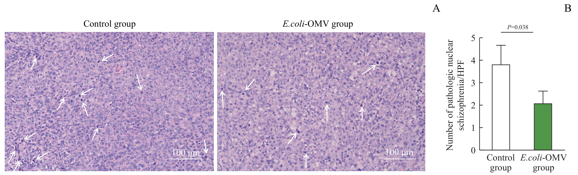
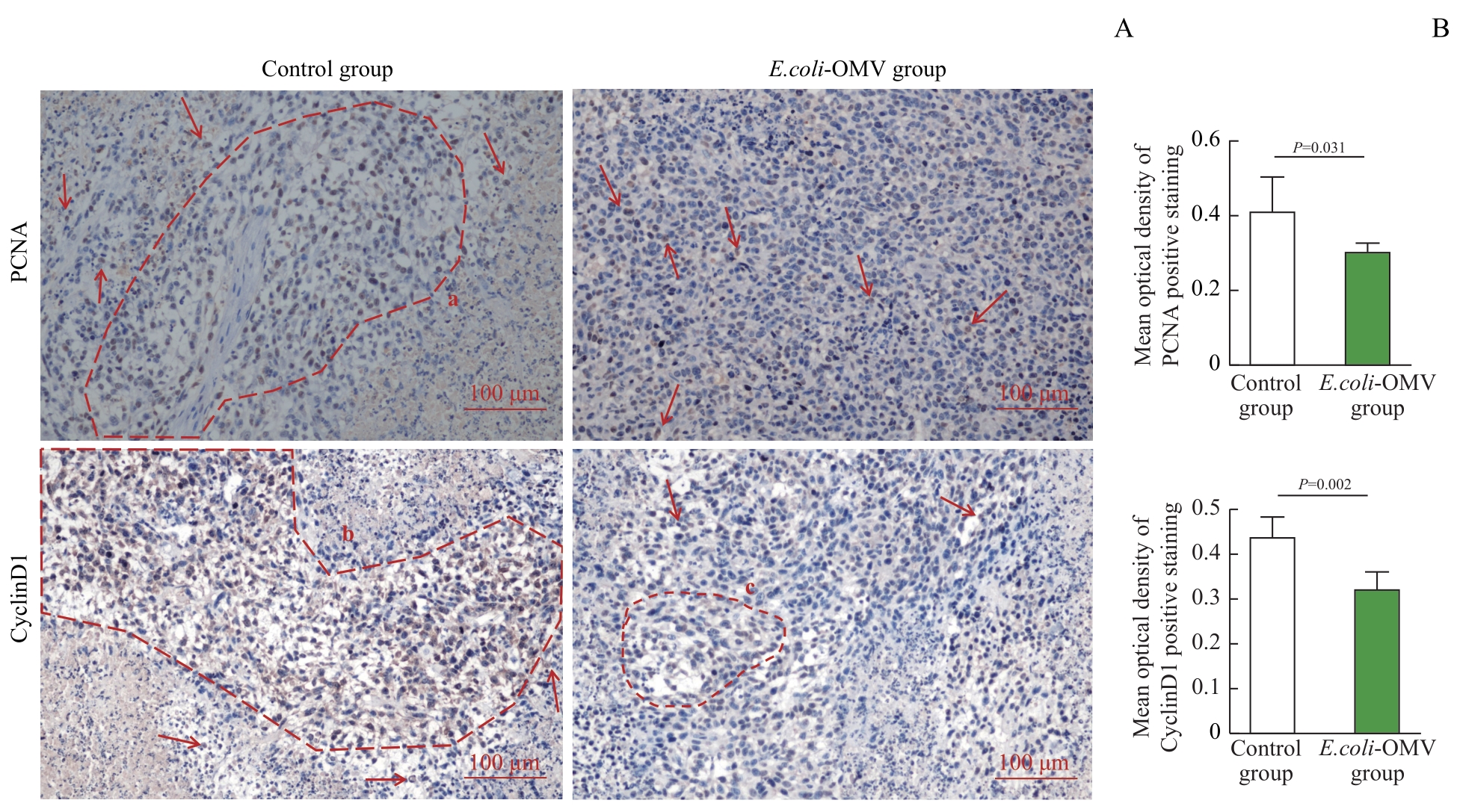
|
|
WANG M, SU W Y, et al. Bacteria: a new weapon against cancer[J]. World Latest Medicine Information, 2019, 19(66): 102-103, 105.
|
| 7 |
SEDIGHI M, ZAHEDI BIALVAEI A, HAMBLIN M R, et al. Therapeutic bacteria to combat cancer; current advances, challenges, and opportunities[J]. Cancer Med, 2019, 8(6): 3167-3181.
|
| 8 |
KIKUCHI Y, OBANA N, TOYOFUKU M, et al. Diversity of physical properties of bacterial extracellular membrane vesicles revealed through atomic force microscopy phase imaging[J]. Nanoscale, 2020, 12(14): 7950-7959.
|
| 9 |
邱晓涵, 李泳江, 吴军勇, 等. 细菌外膜囊泡: 疾病治疗的新途径[J]. 药学学报, 2021, 56(12): 3441-3450.
|
|
QIU X H, LI Y J, WU J Y, et al. Bacterial outer membrane vesicles: a new approach to diseases therapy[J]. Acta Pharmaceutica Sinica, 2021, 56(12): 3441-3450.
|
| 10 |
CHEN Q, BAI H Z, WU W T, et al. Bioengineering bacterial vesicle-coated polymeric nanomedicine for enhanced cancer immunotherapy and metastasis prevention[J]. Nano Lett, 2020, 20(1): 11-21.
|
| 11 |
CHEN Y, LIU L G, FU H, et al. Comparative proteomic analysis of outer membrane vesicles from Shigella flexneri under different culture conditions[J]. Biochem Biophys Res Commun, 2014, 453(4): 696-702.
|
| 12 |
TOYOFUKU M, NOMURA N, EBERL L. Types and origins of bacterial membrane vesicles[J]. Nat Rev Microbiol, 2019, 17(1): 13-24.
|
| 13 |
RUDNICKA M, NOSZCZYŃSKA M, MALICKA M, et al. Outer membrane vesicles as mediators of plant-bacterial interactions[J]. Front Microbiol, 2022, 13: 902181.
|
| 14 |
胡慧冰, 侯昕宇, 贺牧野, 等. 细菌外膜囊泡包覆的载药纳米粒的制备及其小鼠鼻腔免疫效果评价[J]. 上海交通大学学报(医学版), 2018, 38(2): 155-160.
|
|
HU H B, HOU X Y, HE M Y, et al. Preparation of bacterial outer membrane vesicle coated nanoparticle loaded with drug and evaluation of its nasal immune effect in mice[J]. Journal of Shanghai Jiao Tong University (Medical Science), 2018, 38(2): 155-160.
|
| 15 |
YAGHOUBI A, KHAZAEI M, HASANIAN S, et al. Bacteriotherapy in breast cancer[J]. Int J Mol Sci, 2019, 20(23): 5880.
|
| 16 |
FARKAS-HIMSLEY H, CHEUNG R. Bacterial proteinaceous products (bacteriocins) as cytotoxic agents of neoplasia[J]. Cancer Res, 1976, 36(10): 3561-3567.
|
| 17 |
STRZALKA W, ZIEMIENOWICZ A. Proliferating cell nuclear antigen (PCNA): a key factor in DNA replication and cell cycle regulation[J]. Ann Bot, 2011, 107(7): 1127-1140.
|
| 18 |
GOLIAS C H, CHARALABOPOULOS A, CHARALABOPOULOS K. Cell proliferation and cell cycle control: a mini review[J]. Int J Clin Pract, 2004, 58(12): 1134-1141.
|
 ), 马官荣1,2, 姜咏竹1,2, 常秀林1,2, 方廖琼1, 白晋1,2(