上海交通大学学报(医学版) ›› 2024, Vol. 44 ›› Issue (8): 999-1010.doi: 10.3969/j.issn.1674-8115.2024.08.009

• 论著 · 基础研究 • 上一篇    下一篇

PRMT6促进乳腺癌细胞的增殖和迁移

韩依杉1(), 徐梓淇1, 陶梦玉1, 范广建1, 余波2,3()   

  1. 1.上海交通大学医学院附属第一人民医院肿瘤中心,上海 201600
    2.复旦大学附属肿瘤医院肿瘤内科,上海 200032
    3.复旦大学上海医学院肿瘤学系,上海 200032
  • 收稿日期:2024-03-03 接受日期:2024-04-19 出版日期:2024-08-28 发布日期:2024-08-27
  • 通讯作者: 余波 E-mail:hanyishan97@163.com;yudky@163.com
  • 作者简介:韩依杉(1997—),女,硕士生;电子信箱:hanyishan97@163.com
  • 基金资助:
    国家自然科学基金(82103261);上海市“科技创新行动计划”自然科学基金(23ZR1412300)

PRMT6 promotes the proliferation and migration of breast cancer cells

HAN Yishan1(), XU Ziqi1, TAO Mengyu1, FAN Guangjian1, YU Bo2,3()   

  1. 1.Department of Oncology Center, Shanghai General Hospital, Shanghai Jiao Tong University School of Medicine, Shanghai 201600, China
    2.Department of Medical Oncology, Fudan University Shanghai Cancer Center, Shanghai 200032, China
    3.Department of Oncology, Shanghai Medical College, Fudan University, Shanghai 200032, China.
  • Received:2024-03-03 Accepted:2024-04-19 Online:2024-08-28 Published:2024-08-27
  • Contact: YU Bo E-mail:hanyishan97@163.com;yudky@163.com
  • Supported by:
    National Natural Science Foundation of China(82103261);Foundation of Shanghai Municipal Science and Technology Commission(23ZR1412300)

摘要:

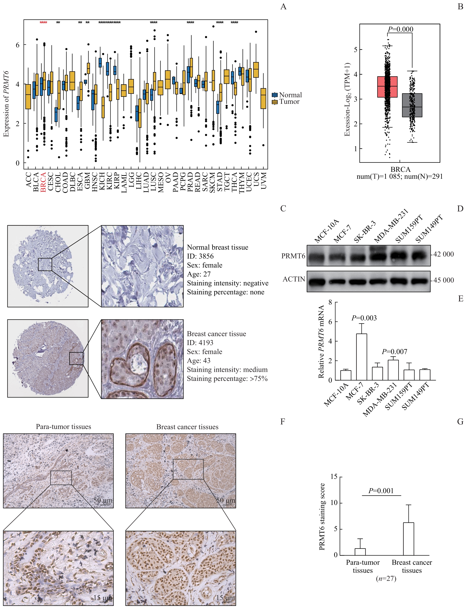
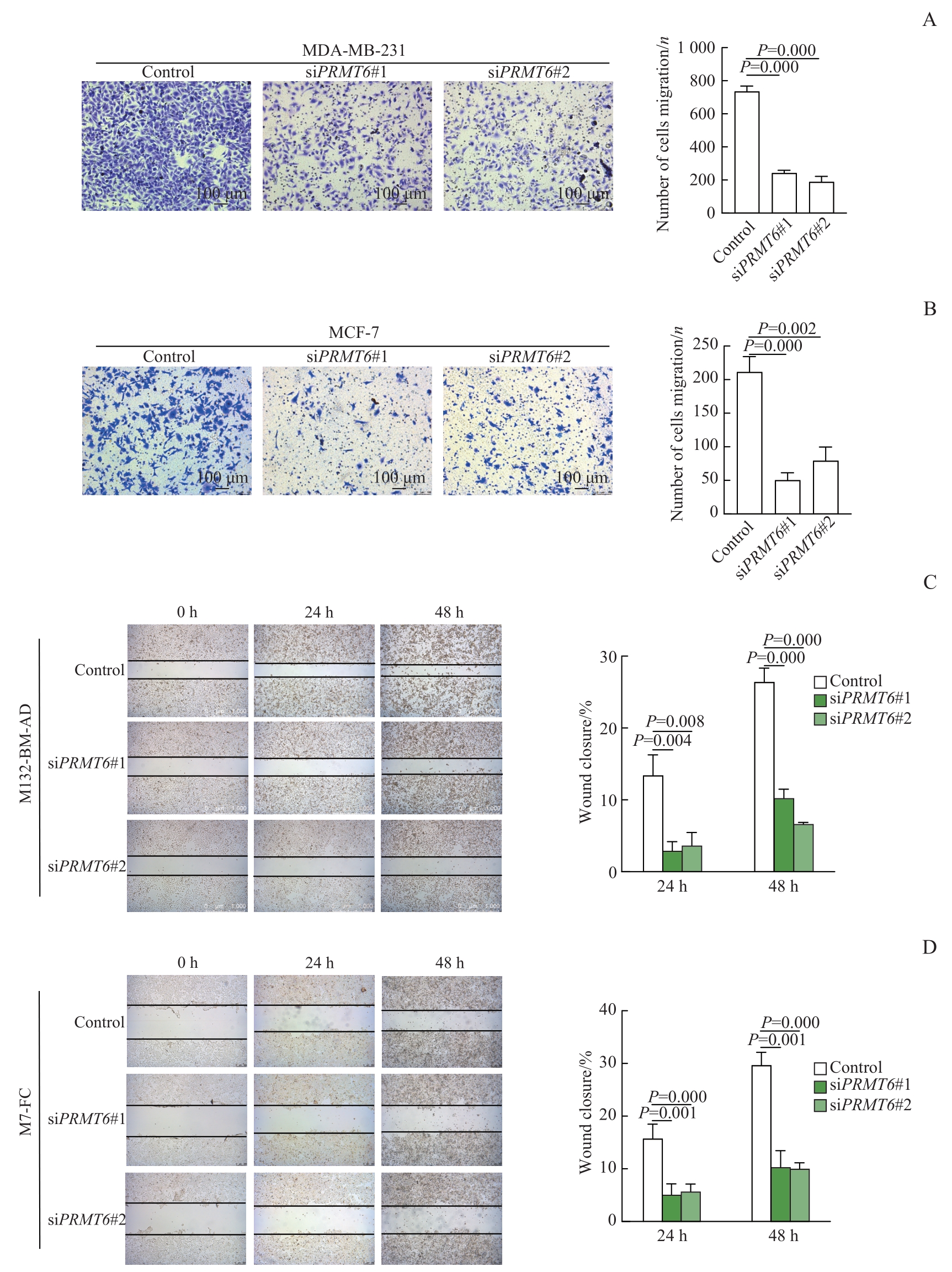
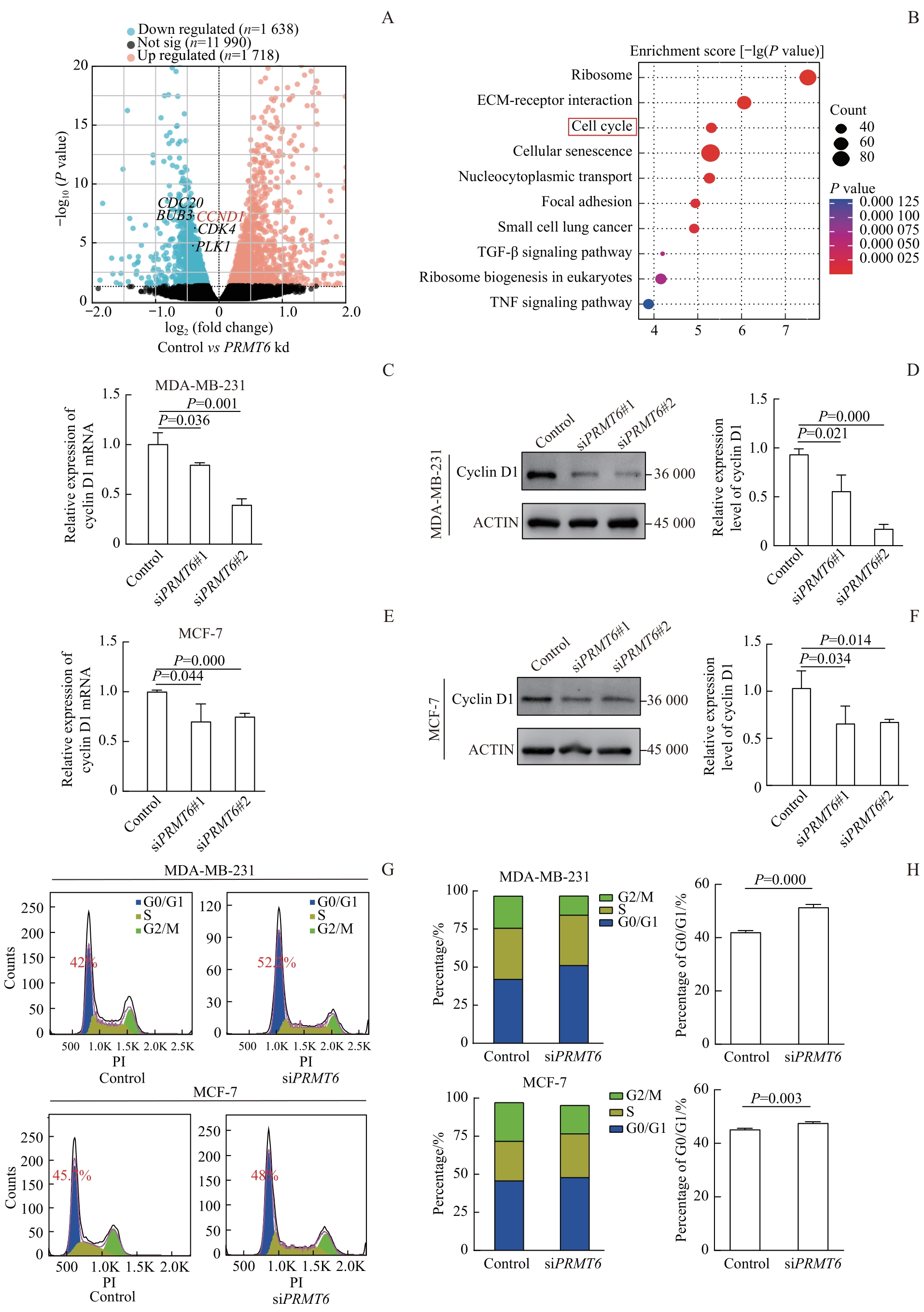
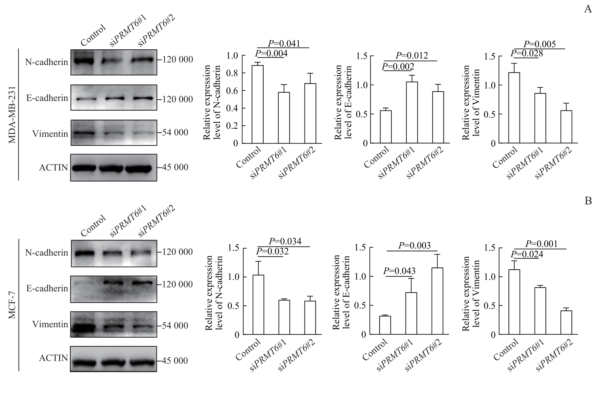
目的·研究蛋白质精氨酸甲基转移酶6(protein arginine methyltransferase 6,PRMT6)在乳腺癌中的表达及其对乳腺癌细胞增殖和迁移能力的影响。方法·通过R语言分析癌症基因组图谱(The Cancer Genome Atlas,TCGA)数据库中PRMT6 mRNA在多种癌症中的表达情况。采用基因表达谱交互分析(Gene Expression Profiling Interactive Analysis,GEPIA2)在线数据库分析PRMT6在正常乳腺组织和乳腺癌组织中的表达差异。利用人类蛋白质图谱(The Human Protein Atlas,HPA)数据库获得人正常乳腺组织和乳腺癌组织的免疫组织化学数据,分析PRMT6的蛋白表达情况。使用免疫组织化学法(immunohistochemistry,IHC)检测27例乳腺癌组织及配对癌旁组织中PRMT6的表达。通过小干扰RNA(small interfering RNA,siRNA)转染技术在MDA-MB-231和MCF-7细胞系中敲低PRMT6,实时荧光定量PCR(quantitative real-time PCR,qRT-PCR)及Western blotting在转录和蛋白水平验证PRMT6的敲低效率。通过细胞计数试剂盒8(cell counting kit-8,CCK-8)、克隆形成实验探究PRMT6对乳腺癌细胞增殖能力的影响。通过细胞划痕实验和Transwell实验探究PRMT6对乳腺癌细胞迁移能力的影响。利用基因表达综合(Gene Expression Omnibus,GEO)数据库中GSE210948数据集的转录组测序数据分析对照组和PRMT6低表达组的差异基因,并进行京都基因与基因组数据库(Kyoto Encyclopedia of Genes and Genomes,KEGG)通路富集分析。使用流式细胞术进行细胞周期分析。采用Western blotting技术检测增殖和迁移相关靶蛋白细胞周期蛋白D1(cyclin D1)、E-钙黏蛋白(E-cadherin)、N-钙黏蛋白(N-cadherin)和波形蛋白(Vimentin)的表达。结果·生物信息学相关分析显示,PRMT6在乳腺癌组织中的表达高于正常乳腺组织(P=0.000)。IHC结果显示,PRMT6在乳腺癌组织中的表达显著高于配对癌旁组织(P=0.001)。qRT-PCR及Western blotting验证MDA-MB-231和MCF-7细胞系中PRMT6的mRNA及蛋白质表达水平,发现与对照组相比,siRNA(siPRMT6#1、siPRMT6#2)显著降低两种细胞中PRMT6 mRNA(P=0.006,P=0.004;P=0.001,P=0.043)和蛋白(P=0.035,P=0.001;P=0.003,P=0.002)的表达水平。敲低PRMT6显著降低MDA-MB-231和MCF-7细胞的增殖(P=0.014,P=0.000;P=0.003,P=0.003)和迁移能力(P=0.000,P=0.000;P=0.000,P=0.002)。KEGG通路富集分析显示,PRMT6的表达影响细胞周期通路。Western blotting结果显示,敲低PRMT6后,与细胞周期通路相关的cyclin D1蛋白水平下降(P=0.021,P=0.000;P=0.034,P=0.014);qRT-PCR结果显示,敲低PRMT6后,cyclin D1转录水平明显下降(P=0.036,P=0.001;P=0.044,P=0.000)。流式细胞术结果显示,敲低PRMT6后,G0/G1期细胞增加(P=0.000;P=0.003),G2/M期细胞减少。下调PRMT6表达后,与细胞迁移相关的E-cadherin表达增加(P=0.002,P=0.012;P=0.043,P=0.003),N-cadherin(P=0.004,P=0.041;P=0.032,P=0.034)和Vimentin(P=0.028,P=0.005;P=0.024,P=0.001)的蛋白表达减少。结论·PRMT6在乳腺癌组织中表达升高,可促进乳腺癌的增殖和迁移。

关键词: 乳腺癌, 蛋白质精氨酸甲基转移酶6, 增殖, 迁移, 细胞周期

Abstract:

Objective ·To examine the expression level of protein arginine methyltransferase 6 (PRMT6) in breast carcinoma tissues and to assess its impact on the proliferative and migratory behaviors of breast cancer cells. Methods ·The PRMT6 transcriptome sequencing data between 33 tumor tissues and normal tissues from The Cancer Genome Atlas (TCGA) database was analyzed through the R language. The gene expression profile interactive analysis (GEPIA2) online database was used to analyze the difference of PRMT6 expression in normal breast tissues and breast cancer tissues. By using the immunohistochemistry (IHC) data of human normal breast tissues and breast cancer tissues from Human Protein Atlas (HPA) database to analyze the protein expression of PRMT6. IHC was used to detect the expression of PRMT6 in breast cancer tissues and paired para-tumor tissues from 27 clinical samples. After PRMT6 was knocked down with small interfering RNA (siRNA) in MDA-MB-231 and MCF-7 cells, the expression of PRMT6 was detected by qRT-PCR and Western blotting. The proliferation ability of breast cancer cells was measured with cell counting kit-8 (CCK-8) assay and colony formation assay. The effect of PRMT6 on the migration ability of breast cancer cells was detected by wound healing assay and Transwell assay. By using the RNA-sequence data from GSE210948 of Gene Expression Omnibus (GEO) database, differentially expressed genes were analyzed in control and low expression groups of PRMT6. Kyoto Encyclopedia of Genes and Genomes (KEGG) pathway enrichment analysis was performed to reveal the signaling pathways associated with PRMT6. Cell cycle analysis was detected by flow cytometry. The expressions of cyclin D1 and EMT-related proteins (E-cadherin, N-cadherin and Vimentin) were detected by Western blotting after knocking down PRMT6. Results ·Bioinformatics analysis and IHC results showed that PRMT6 was highly expressed in breast cancer tissues compared with normal tissues (P=0.000) and para-tumor tissues (P=0.001). qRT-PCR and Western blotting results verified that the siRNA significantly reduced the expression level of PRMT6 in MDA-MB-231 and MCF-7 cell lines compared with the control group (mRNA: P=0.006, P=0.004; P=0.001, P=0.043. Protein: P=0.035, P=0.001; P=0.003, P=0.002). After knocking down PRMT6, the proliferation (P=0.014, P=0.000; P=0.003, P=0.003) and migration (P=0.000, P=0.000; P=0.000, P=0.002) ability of breast cancer cells were inhibited significantly. The KEGG pathway enrichment analysis showed that the expression of PRMT6 affected the cell cycle pathway. After knocking down PRMT6, the expression of cyclin D1 decreased in protein level (P=0.021, P=0.000; P=0.034, P=0.014) and transcription level (P=0.036, P=0.001; P=0.044, P=0.000). Knock down of PRMT6 increased the number of cells in G0/G1 phase (P=0.000; P=0.003) and decreased the number of cells in G2/M phase of the cell cycle. The expression level of E-cadherin increased (P=0.002, P=0.012; P=0.043, P=0.003), while the expression levels of N-cadherin (P=0.004, P=0.041; P=0.032, P=0.034) and Vimentin (P=0.028, P=0.005; P=0.024, P=0.001) decreased in PRMT6 knockdown cells. Conclusion ·PRMT6 is highly expressed in breast cancer, which can promote the proliferation and migration of breast cancer cells.

Key words: breast cancer, protein arginine methyltransferase 6, proliferation, migration, cell cycle

中图分类号: