| [1] |
ZHOU L, CUI C L, LIAN B, et al. Postoperative radiotherapy in resected sinonasal mucosal melanoma[J]. J Clin Oncol, 2019, 37(15_suppl): e21059.
|
| [2] |
SALEH M, JAVADI S, ELSHERIF S, et al. Multimodality imaging and genetics of primary mucosal melanomas and response to treatment[J]. Radiographics, 2021, 41(7): 1954-1972.
|
| [3] |
OTTAVIANO M, GIUNTA E F, MARANDINO L, et al. Anorectal and genital mucosal melanoma: diagnostic challenges, current knowledge and therapeutic opportunities of rare melanomas[J]. Biomedicines, 2022, 10(1): 150.
|
| [4] |
KOTTSCHADE L A, POND G R, OLSZANSKI A J, et al. SALVO: single-arm trial of ipilimumab and nivolumab as adjuvant therapy for resected mucosal melanoma[J]. Clin Cancer Res, 2023, 29(12): 2220-2225.
|
| [5] |
SI L, FANG M Y, CHEN Y, et al. Atezolizumab in combination with bevacizumab in patients with unresectable locally advanced or metastatic mucosal melanoma: interim analysis of an open-label phase II trial[J]. J Clin Oncol, 2021, 39(15_suppl): 9511.
|
| [6] |
JOHNSON D B, SWETTER S M, SALAMA A K S, et al. Cutaneous melanoma: management of melanoma brain metastases and molecular testing[J]. J Natl Compr Cancer Netw, 19(5.5): 644-647.
|
| [7] |
LI S M, WU X W, YAN X Q, et al. Toripalimab plus axitinib in patients with metastatic mucosal melanoma: 3-year survival update and biomarker analysis[J]. J Immunother Cancer, 2022, 10(2): e004036.
|
| [8] |
BLACKBURN S D, SHIN H, FREEMAN G J, et al. Selective expansion of a subset of exhausted CD8 T cells by alphaPD-L1 blockade[J]. Proc Natl Acad Sci USA, 2008, 105(39): 15016-15021.
|
| [9] |
ZENG S S, HU H L, LI Z Y, et al. Local TSH/TSHR signaling promotes CD8+ T cell exhaustion and immune evasion in colorectal carcinoma[J]. Cancer Commun (Lond), 2024, 44(11): 1287-1310.
|
| [10] |
GAO Y, OUYANG Z J, YANG C, et al. Overcoming T cell exhaustion via immune checkpoint modulation with a dendrimer-based hybrid nano complex[J]. Adv Healthc Mater, 2021, 10(19): e2100833.
|
| [11] |
BELK J A, DANIEL B, SATPATHY A T. Epigenetic regulation of T cell exhaustion[J]. Nat Immunol, 2022, 23(6): 848-860.
|
| [12] |
KANG K, LIN X, CHEN P, et al. T cell exhaustion in human cancers[J]. Biochim Biophys Acta Rev Cancer, 2024, 1879(5): 189162.
|
| [13] |
MILLER B C, SEN D R, AL ABOSY R, et al. Subsets of exhausted CD8+ T cells differentially mediate tumor control and respond to checkpoint blockade[J]. Nat Immunol, 2019, 20(3): 326-336.
|
| [14] |
KALTENMEIER C, YAZDANI H O, MORDER K, et al. Neutrophil extracellular traps promote T cell exhaustion in the tumor microenvironment[J]. Front Immunol, 2021, 12: 785222.
|
| [15] |
GIESE M A, HIND L E, HUTTENLOCHER A. Neutrophil plasticity in the tumor microenvironment[J]. Blood, 2019, 133(20): 2159-2167.
|
| [16] |
WANG L W, LIU Y H, DAI Y T, et al. Single-cell RNA-seq analysis reveals BHLHE40-driven pro-tumour neutrophils with hyperactivated glycolysis in pancreatic tumour microenvironment[J]. Gut, 2023, 72(5): 958-971.
|
| [17] |
MENG Y, YE F, NIE P P, et al. Immunosuppressive CD10+ALPL+ neutrophils promote resistance to anti-PD-1 therapy in HCC by mediating irreversible exhaustion of T cells[J]. J Hepatol, 2023, 79(6): 1435-1449.
|
| [18] |
HUANG C T, LIAO Y H, LIAU J Y, et al. Pretreatment neutrophil-to-lymphocyte ratio predicts survival in primary mucosal melanoma[J]. J Am Acad Dermatol, 2025, 92(2): 340-343.
|
| [19] |
SADE-FELDMAN M, YIZHAK K, BJORGAARD S L, et al. Defining T cell states associated with response to checkpoint immunotherapy in melanoma[J]. Cell, 2018, 175(4): 998-1013.e20.
|
| [20] |
TIROSH I, IZAR B, PRAKADAN S M, et al. Dissecting the multicellular ecosystem of metastatic melanoma by single-cell RNA-seq[J]. Science, 2016, 352(6282): 189-196.
|
| [21] |
ZHANG C, SHEN H R, YANG T L, et al. A single-cell analysis reveals tumor heterogeneity and immune environment of acral melanoma[J]. Nat Commun, 2022, 13(1): 7250.
|
| [22] |
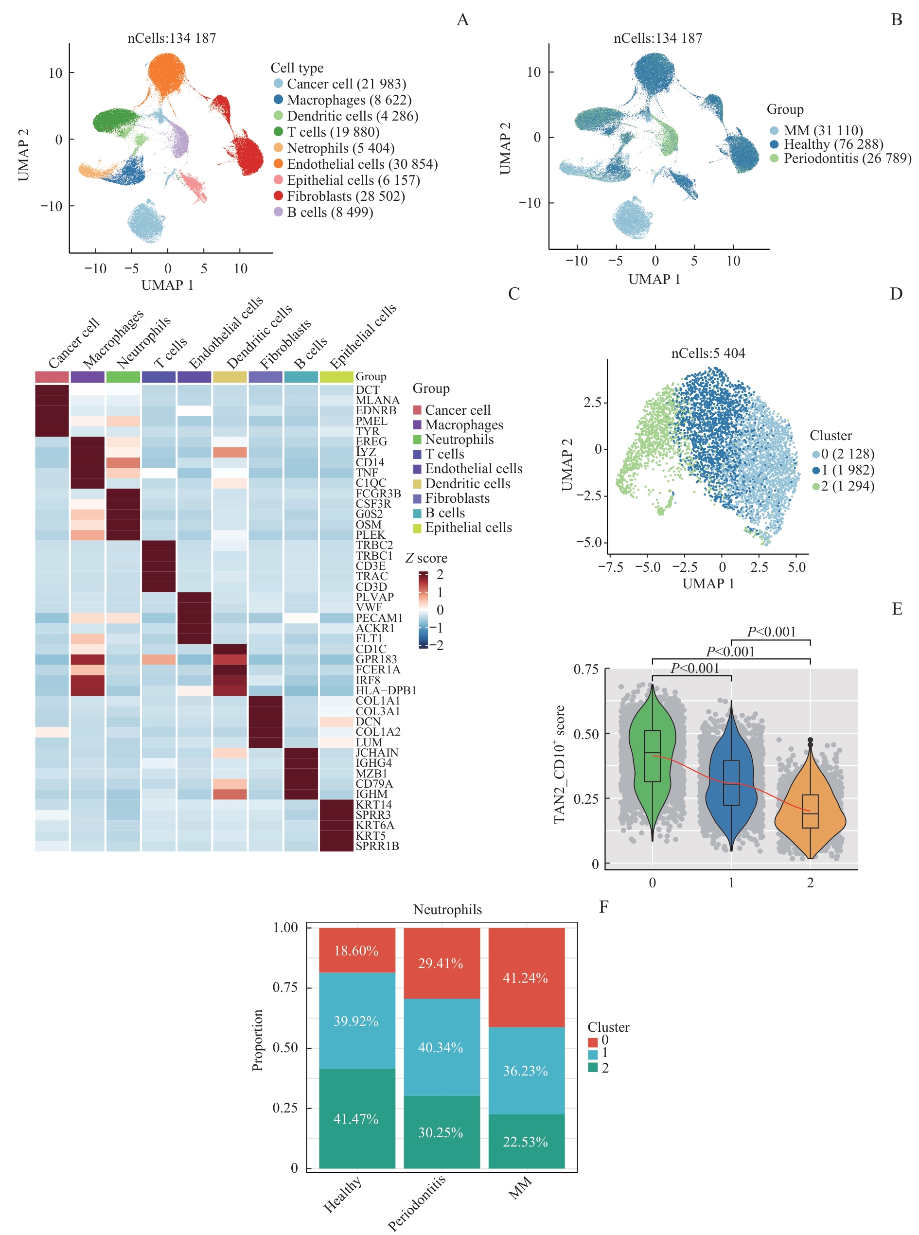
ZHOU Z Y, ZHOU X X, JIANG X, et al. Single-cell profiling identifies IL1Bhi macrophages associated with inflammation in PD-1 inhibitor-induced inflammatory arthritis[J]. Nat Commun, 2024, 15(1): 2107.
|
| [23] |
ALSHETAIWI H, PERVOLARAKIS N, MCINTYRE L L, et al. Defining the emergence of myeloid-derived suppressor cells in breast cancer using single-cell transcriptomics[J]. Sci Immunol, 2020, 5(44): eaay6017.
|
| [24] |
KELLIHER M A, RODERICK J E. NOTCH signaling in T-cell-mediated anti-tumor immunity and T-cell-based immunotherapies[J]. Front Immunol, 2018, 9: 1718.
|
| [25] |
LIU W, STACHURA P, XU H C, et al. BAFF attenuates immunosuppressive monocytes in the melanoma tumor microenvironment[J]. Cancer Res, 2022, 82(2): 264-277.
|
| [26] |
LIU X B, WANG Y Y, BAUER A T, et al. Neutrophils activated by membrane attack complexes increase the permeability of melanoma blood vessels[J]. Proc Natl Acad Sci USA, 2022, 119(33): e2122716119.
|
| [27] |
BARON M, IDEKER T. Abstract 178: desmosome mutations in melanoma promote cellular proliferation and disease progression[J]. Cancer Res, 2021, 81(13_Supplement): 178.
|
| [28] |
IRIONDO O, YU M. Unexpected friendship: neutrophils help tumor cells en route to metastasis[J]. Dev Cell, 2019, 49(3): 308-310.
|
| [29] |
GUNGABEESOON J, GORT-FREITAS N A, KISS M, et al. A neutrophil response linked to tumor control in immunotherapy[J]. Cell, 2023, 186(7): 1448-1464.e20.
|
| [30] |
WU Y C, MA J Q, YANG X P, et al. Neutrophil profiling illuminates anti-tumor antigen-presenting potency[J]. Cell, 2024, 187(6): 1422-1439.e24.
|
| [31] |
LIU S J, YUAN S J, LIU M C, et al. In situ tumor cell engineering reverses immune escape to enhance immunotherapy effect[J]. Acta Pharm Sin B, 2025, 15(1): 627-641.
|
| [32] |
BRANDAU S, HARTL D. Lost in neutrophil heterogeneity? CD10![J]. Blood, 2017, 129(10): 1240-1241.
|
| [33] |
SÁNCHEZ-MAGRANER L, MILES J, BAKER C L, et al. High PD-1/PD-L1 checkpoint interaction infers tumor selection and therapeutic sensitivity to anti-PD-1/PD-L1 treatment[J]. Cancer Res, 2020, 80(19): 4244-4257.
|
| [34] |
MA H Y, YAMAMOTO G, XU J, et al. IL-17 signaling in steatotic hepatocytes and macrophages promotes hepatocellular carcinoma in alcohol-related liver disease[J]. J Hepatol, 2020, 72(5): 946-959.
|
| [35] |
HAO Y W, LIU D L, WANG K Y, et al. Imaging and therapy of tumors based on neutrophil extracellular traps[J]. Small Sci, 2024, 4(10): 2400212.
|
| [36] |
YANG L Y, LUO Q, LU L, et al. Increased neutrophil extracellular traps promote metastasis potential of hepatocellular carcinoma via provoking tumorous inflammatory response[J]. J Hematol Oncol, 2020, 13(1): 3.
|
| [37] |
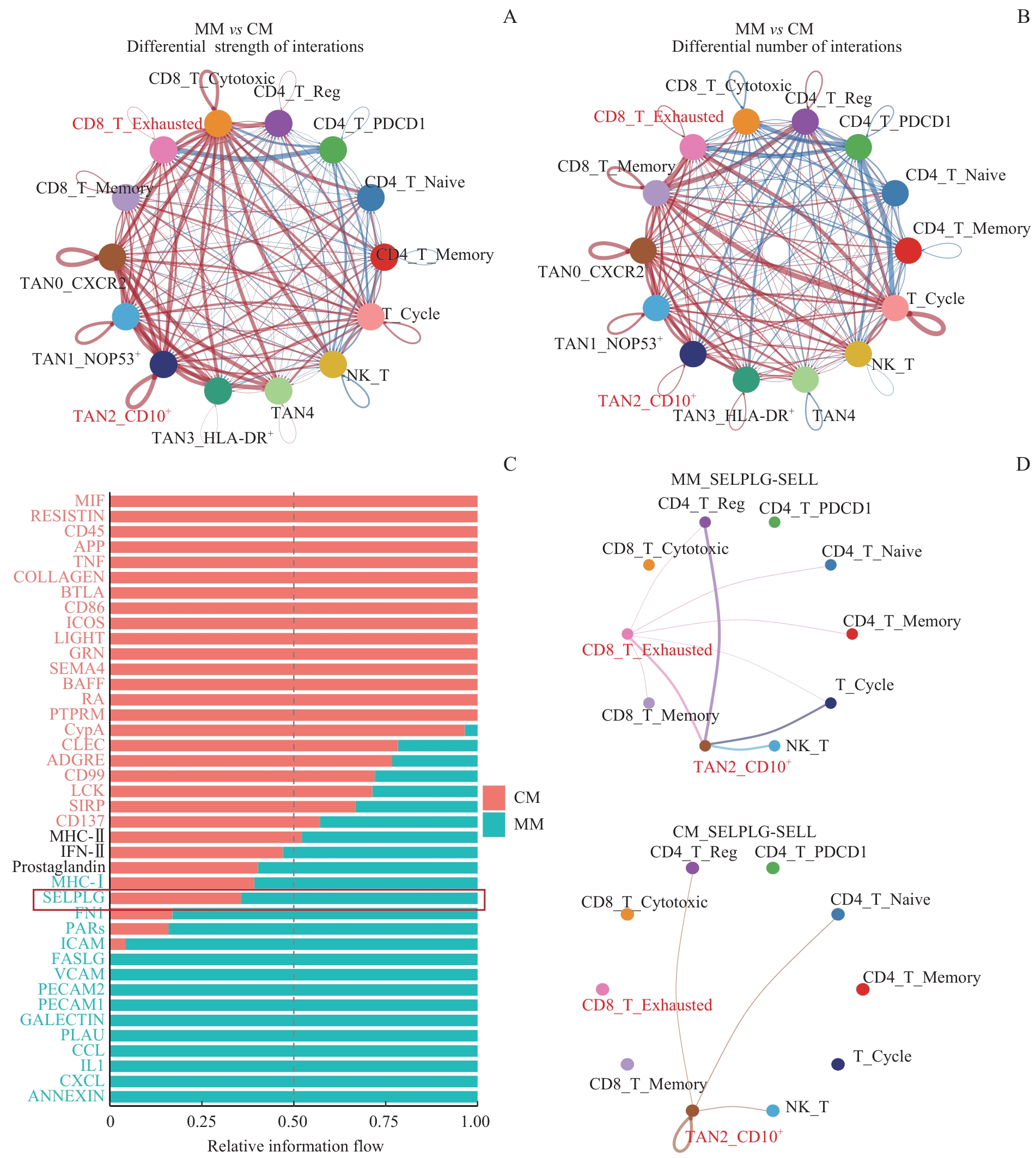
VIRAMONTES K M, NEUBERT E N, DEROGATIS J M, et al. PD-1 immune checkpoint blockade and PSGL-1 inhibition synergize to reinvigorate exhausted T cells[J]. Front Immunol, 2022, 13: 869768.
|
| [38] |
van NOT O J, van DEN EERTWEGH A J M, JALVING H, et al. Long-term survival in patients with advanced melanoma[J]. JAMA Netw Open, 2024, 7(8): e2426641.
|
| [39] |
de MEZA M M, van NOT O J, BLOKX W, et al. Efficacy of checkpoint inhibition in advanced acral melanoma[J]. J Clin Oncol, 2021, 39(15_suppl): e21527.
|
| [40] |
ZENG W Y, WANG Y, ZHANG Q, et al. Neutrophil nanodecoys inhibit tumor metastasis by blocking the interaction between tumor cells and neutrophils[J]. ACS Nano, 2024, 18(10): 7363-7378.
|
| [41] |
QUE H Y, FU Q M, LAN T X, et al. Tumor-associated neutrophils and neutrophil-targeted cancer therapies[J]. Biochim Biophys Acta Rev Cancer, 2022, 1877(5): 188762.
|
 )
)