
上海交通大学学报(医学版) ›› 2026, Vol. 46 ›› Issue (8): 997-1006.doi: 10.3969/j.issn.1674-8115.2026.08.001
• 前沿述评 • 下一篇
杨蕊馨, 于颖彦( )
)
收稿日期:2026-01-09
接受日期:2026-04-08
出版日期:2026-08-28
发布日期:2026-08-28
通讯作者:
于颖彦,教授,博士;电子信箱:ruijinhospitalyyy@163.com。作者简介:第一联系人:杨蕊馨撰写并修改论文,于颖彦审阅论文。所有作者均阅读并同意最终稿件的提交。
基金资助:
Yang Ruixin, Yu Yingyan()
Received:2026-01-09
Accepted:2026-04-08
Online:2026-08-28
Published:2026-08-28
Contact:
Yu Yingyan, E-mail: ruijinhospitalyyy@163.com.About author:First author contact:The manuscript was drafted and revised by Yang Ruixin, and reviewed by Yu Yingyan. Both authors have read the final version of paper and consented to its submission.
Supported by:摘要:
类器官作为新型体外三维培养模型,凭借其高度自组织能力,可以很好地还原其来源组织的形态结构、病理生理功能、基因变异谱系以及药物治疗反应性。与传统的动物模型相比,类器官具有制备周期短、成本低、伦理争议小和通量高等特点,目前被广泛应用于生物医药研究。患者源性类器官模型保留了其来源组织的异质性,可以体外长期扩增传代培养,且低温冻存复苏后依然保持细胞活力,这为人类疾病建模提供了宝贵的平台,并在发病机制探索、新型药物筛选及药物敏感性预测中展现出重要的应用价值。而人工智能(artificial intelligence,AI)的介入加速了类器官构建与应用的自动化与标准化进程。该文系统总结了消化系统良恶性疾病相关类器官研究进展,尤其是不同器官组织源性类器官构建的优化方案,并就消化系统肿瘤样本采集方法、新辅助治疗对类器官构建的影响等进行分析。此外,文章还概述了AI算法在类器官构建及应用中的进展,包括AI辅助的类器官形态表征、细胞活力评估、药物敏感性预测,以及整合AI的自动化类器官平台建设现状。展望未来,AI将在消化系统疾病类器官构建与多种应用场景中发挥重要作用,为疾病建模、分子靶点发现、药物筛选、耐药机制解析等转化应用提供强有力的支撑。
中图分类号:
杨蕊馨, 于颖彦. 消化系统疾病类器官的构建应用及其与人工智能整合的研究进展[J]. 上海交通大学学报(医学版), 2026, 46(8): 997-1006.
Yang Ruixin, Yu Yingyan. Research progress in the construction and application of digestive system disease organoids and their integration with artificial intelligence[J]. Journal of Shanghai Jiao Tong University (Medical Science), 2026,(8): 997-1006.
| Site | Biomarker | Differential diagnosis |
|---|---|---|
| Esophagus | KRT5, KRT14 | Squamous epithelium: KRT5 Glandular epithelium (such as Barrett esophagus): CDX2, MUC2, KRT20 |
| Stomach | TFF1, MUC5AC, MUC6, MUC2, KRT8 | Surface epithelium: TFF1 Fundic and corpus gland: MUC6, PGC, ATP4A Basal stem cell: LGR5 Endocrine cell: CHGA, CHGB, SCG5 |
| Intestine | EPCAM, KRT20, CDX2 | Duodenum: SLC5A1, CYP3A4 Jejunum: SLC6A19, LCT Ileum: IL22RA1, SLC10A2 Colon: CA2, MUC4 |
| Liver and bile duct | KRT7, KRT19 | Liver: ALB, AAT, TTR, APOA2 Bile duct: KRT7, KRT19, SOX9 |
| Pancreas | Endocrine and exocrine glands have different marker genes | Exocrine gland: PRSS1, CPA1, KRT19 Endocrine gland: CHGA, INS, GCG |
表1 消化系统不同部位上皮组织类器官的细胞鉴定标志物
Tab 1 Cell identification markers for organoids derived from different sites of the digestive system
| Site | Biomarker | Differential diagnosis |
|---|---|---|
| Esophagus | KRT5, KRT14 | Squamous epithelium: KRT5 Glandular epithelium (such as Barrett esophagus): CDX2, MUC2, KRT20 |
| Stomach | TFF1, MUC5AC, MUC6, MUC2, KRT8 | Surface epithelium: TFF1 Fundic and corpus gland: MUC6, PGC, ATP4A Basal stem cell: LGR5 Endocrine cell: CHGA, CHGB, SCG5 |
| Intestine | EPCAM, KRT20, CDX2 | Duodenum: SLC5A1, CYP3A4 Jejunum: SLC6A19, LCT Ileum: IL22RA1, SLC10A2 Colon: CA2, MUC4 |
| Liver and bile duct | KRT7, KRT19 | Liver: ALB, AAT, TTR, APOA2 Bile duct: KRT7, KRT19, SOX9 |
| Pancreas | Endocrine and exocrine glands have different marker genes | Exocrine gland: PRSS1, CPA1, KRT19 Endocrine gland: CHGA, INS, GCG |
| Sample origin | Sample size | Sample collection requirement | Note |
|---|---|---|---|
| Surgical resection | 200 mg (approximately the size of 2 soybeans) | Selecting vascular-rich, soft tissue; avoiding central necrotic areas tissues with marked fibrosis, large adipose aggregates, hard consistency, electrocoagulation artifacts, or yellowish-gray discoloration | Optimizing enzymatic digestion time for pancreatic cancer organoids to suppress excessive stromal cell growth |
| Needle biopsy | 3 pieces | Maximizing cell retention and minimizing cell damage during sample processing and digestion | Magnifying endoscopy can be employed for sample collection to ensure high tumor purity and avoid necrotic areas in specimens |
| Malignant ascites | 50‒100 mL | Collecting sufficient ascites, harvesting cells via centrifugation, and preparing a single-cell suspension after red blood cell lysis | Excess immunocytes can be removed by Ficoll gradient centrifugation |
表2 消化系统肿瘤类器官构建中肿瘤样本采集注意事项
Tab 2 Precautions for tumor sample collection in the organoid construction of digestive system tumor
| Sample origin | Sample size | Sample collection requirement | Note |
|---|---|---|---|
| Surgical resection | 200 mg (approximately the size of 2 soybeans) | Selecting vascular-rich, soft tissue; avoiding central necrotic areas tissues with marked fibrosis, large adipose aggregates, hard consistency, electrocoagulation artifacts, or yellowish-gray discoloration | Optimizing enzymatic digestion time for pancreatic cancer organoids to suppress excessive stromal cell growth |
| Needle biopsy | 3 pieces | Maximizing cell retention and minimizing cell damage during sample processing and digestion | Magnifying endoscopy can be employed for sample collection to ensure high tumor purity and avoid necrotic areas in specimens |
| Malignant ascites | 50‒100 mL | Collecting sufficient ascites, harvesting cells via centrifugation, and preparing a single-cell suspension after red blood cell lysis | Excess immunocytes can be removed by Ficoll gradient centrifugation |
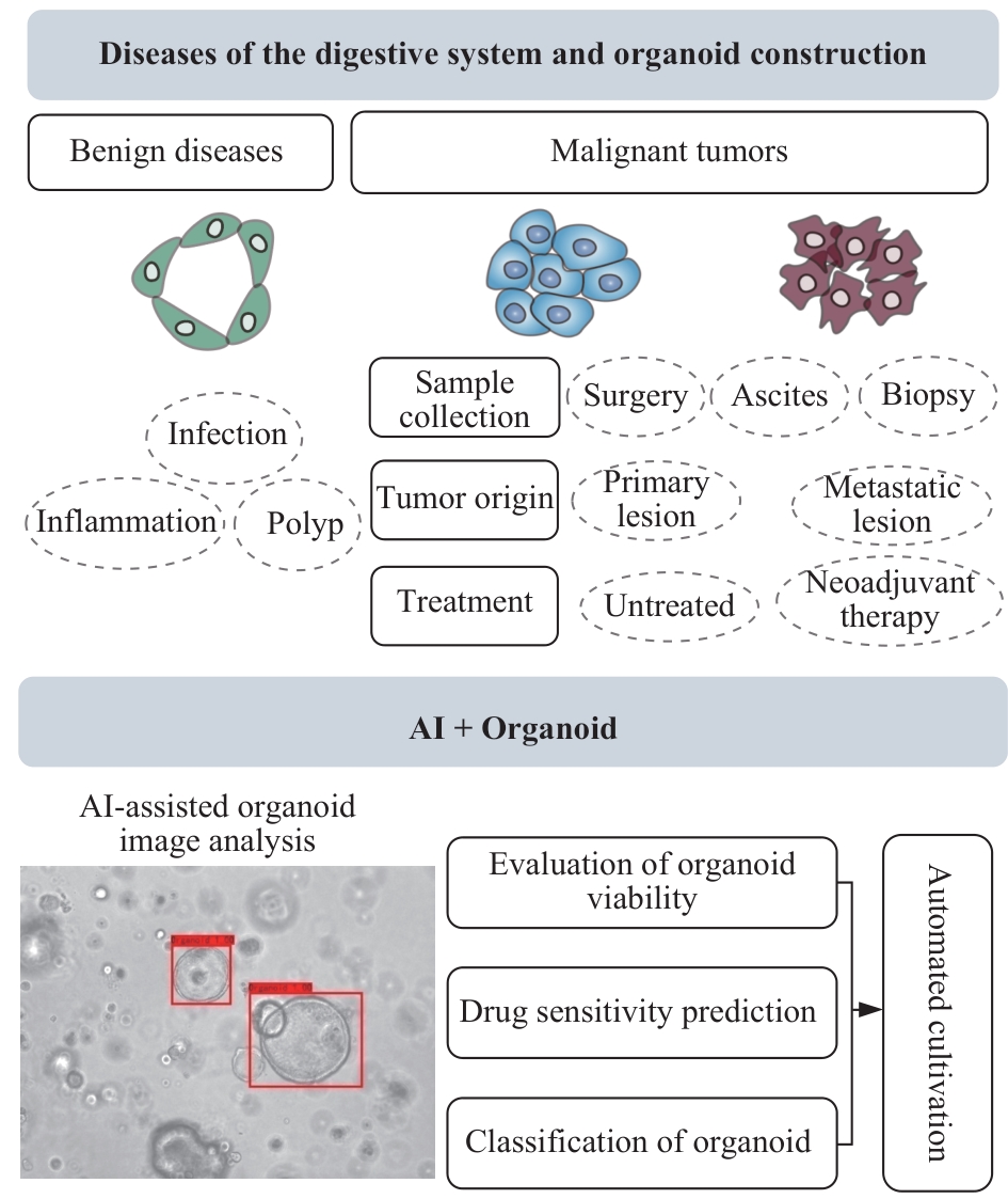
图1 消化系统疾病类器官构建中AI应用示意图Note: Both benign and malignant tissues of the digestive system can be used for organoid construction and research. Malignant tumor samples have diverse origins. AI algorithms can assist in organoid viability evaluation, drug sensitivity prediction, and phenotypic classification.
Fig 1 Schematic diagram of AI applications in organoid construction for digestive system diseases
| [1] | Sato T, Vries R G, Snippert H J, et al. Single Lgr5 stem cells build crypt-villus structures in vitro without a mesenchymal niche[J]. Nature, 2009, 459(7244): 262-265. |
| [2] | Yang R X, Qi Y, Zhang X Y, et al. Living biobank: standardization of organoid construction and challenges[J]. Chin Med J, 2024, 137(24): 3050-3060. |
| [3] | Wadman M. FDA no longer has to require animal testing for new drugs[J]. Science, 2023, 379(6628): 127-128. |
| [4] | Zhou L B, Huang J J, Li C, et al. Organoids and organs-on-chips: recent advances, applications in drug development, and regulatory challenges[J]. Med, 2025, 6(4): 100667. |
| [5] | Elfatimi E, Lekbach Y, Prakash S, et al. Artificial intelligence-, organoid-, and organ-on-chip-powered models to improve pre-clinical animal testing of vaccines and immunotherapeutics: potential, progress, and challenges[J]. Front Artif Intell, 2025, 8: 1681106. |
| [6] | Liu H Y, Wang X L. Esophageal organoids: applications and future prospects[J]. J Mol Med, 2023, 101(8): 931-945. |
| [7] | Jin R U, Xu Y W, Lih T M, et al. SOX2 regulates foregut squamous epithelial homeostasis and is lost during Barrett's esophagus development[J]. J Clin Invest, 2025, 135(16): e190374. |
| [8] | Yang R X, Du Y T, Kwan W, et al. A quick and reliable image-based AI algorithm for evaluating cellular senescence of gastric organoids[J]. Cancer Biol Med, 2023, 20(7): 519-536. |
| [9] | McCracken K W, Catá E M, Crawford C M, et al. Modelling human development and disease in pluripotent stem-cell-derived gastric organoids[J]. Nature, 2014, 516(7531): 400-404. |
| [10] | Son Y S, Kwon Y H, Lee M S, et al. Helicobacter pylori VacA-induced mitochondrial damage in the gastric pit cells of the antrum and therapeutic rescue[J]. Biomaterials, 2025, 314: 122842. |
| [11] | McGowan K P, Delgado E, Keeley T M, et al. Region-specific Wnt signaling responses promote gastric polyp formation in patients with familial adenomatous polyposis[J]. JCI Insight, 2023, 8(24): e174546. |
| [12] | Rodriguez Mier N, Cuyx S, De Boeck K, et al. Correlation between patient-derived intestinal organoids and clinical responses to CFTR modulators in people with cystic fibrosis homozygous for F508del[J]. J Cyst Fibros, 2025, 24(5): 939-945. |
| [13] | Jurickova I, Dreskin B W, Angerman E, et al. Eicosatetraynoic acid regulates profibrotic pathways in an induced pluripotent stem cell-derived macrophage-human intestinal organoid model of Crohn's disease[J]. J Crohns Colitis, 2025,19(2): jjae139. |
| [14] | Pleguezuelos-Manzano C, Puschhof J, van den Brink S, et al. Establishment and culture of human intestinal organoids derived from adult stem cells[J]. Curr Protoc Immunol, 2020, 130(1): e106. |
| [15] | Zhang S X, Du S J, Wang Y Y, et al. Rapid establishment of murine gastrointestinal organoids using mechanical isolation method[J]. Biochem Biophys Res Commun, 2022, 608: 30-38. |
| [16] | Laborde N, Barusseaud A, Quaranta M, et al. Human colonic organoids for understanding early events of familial adenomatous polyposis pathogenesis[J]. J Pathol, 2025, 265(1): 26-40. |
| [17] | Luce E, Messina A, Duclos-Vallée J C. Hepatic organoids as a platform for liver disease modeling and the development of novel therapies[J]. Clin Res Hepatol Gastroenterol, 2025, 49(7): 102647. |
| [18] | Chen Y K, Liu Y J, Chen S M, et al. Liver organoids: a promising three-dimensional model for insights and innovations in tumor progression and precision medicine of liver cancer[J]. Front Immunol, 2023, 14: 1180184. |
| [19] | Lui V C H. Organoids in biliary atresia[J]. World J Pediatr Surg, 2025, 8(2): e001010. |
| [20] | Liu K, Wang Y N, Li J J, et al. Macrophage-augmented organoids recapitulate the complex pathophysiology of viral diseases and enable development of multitarget therapeutics[J]. Nat Biomed Eng, 2025, 9(11): 1848-1868. |
| [21] | Reich M, Spomer L, Klindt C, et al. Downregulation of TGR5 (GPBAR1) in biliary epithelial cells contributes to the pathogenesis of sclerosing cholangitis[J]. J Hepatol, 2021, 75(3): 634-646. |
| [22] | Sampaziotis F, Muraro D, Tysoe O C, et al. Cholangiocyte organoids can repair bile ducts after transplantation in the human liver[J]. Science, 2021, 371(6531): 839-846. |
| [23] | Huang B, Trujillo M A, Fujikura K, et al. Molecular characterization of organoids derived from pancreatic intraductal papillary mucinous neoplasms[J]. J Pathol, 2020, 252(3): 252-262. |
| [24] | Beato F, Reverón D, Dezsi K B, et al. Establishing a living biobank of patient-derived organoids of intraductal papillary mucinous neoplasms of the pancreas[J]. Lab Invest, 2021, 101(2): 204-217. |
| [25] | Wang D Z, Guo Y B, Zhu J C, et al. Hyaluronic acid methacrylate/pancreatic extracellular matrix as a potential 3D printing bioink for constructing islet organoids[J]. Acta Biomater, 2023, 165: 86-101. |
| [26] | Fernández Á, Casamitjana J, Holguín-Horcajo A, et al. A single-cell atlas of the murine pancreatic ductal tree identifies novel cell populations with potential implications in pancreas regeneration and exocrine pathogenesis[J]. Gastroenterology, 2024, 167(5): 944-960.e15. |
| [27] | 王玉蓉, 汪元元, 翁海燕. 胃肠道平滑肌肉瘤临床病理分析3例报告[J]. 诊断学理论与实践, 2024, 23(5): 537-541. |
| Wang Y R, Wang Y Y, Weng H Y. Clinical and pathological analysis of gastrointestinal leiomyosarcoma: report of three cases[J]. Journal of Diagnostics Concepts & Practice, 2024, 23(5): 537-541. | |
| [28] | Mönch D, Thiel J, Dong M, et al. Feasibility of patient-derived 3D gastrointestinal stromal tumour models as alternatives for in vivo mouse models[J]. Int J Mol Sci, 2025, 26(23): 11456. |
| [29] | 苏新程, 林涛, 黄佳烨, 等. 3D类器官在胃肠间质瘤诊治中的研究及临床应用[J]. 临床外科杂志, 2025, 33(6): 568-571. |
| Su X C, Lin T, Huang J Y, et al. Research and clinical application of 3D organoids in the diagnosis and treatment of gastrointestinal stromal tumors[J]. Journal of Clinical Surgery, 2025, 33(6): 568-571. | |
| [30] | 吴琪, 范伯男, 李岩. 2022全球癌症统计报告分析解读: 中国与世界癌症疾病负担与流行趋势[J]. 诊断学理论与实践, 2025, 24(2): 135-145. |
| Wu Q, Fan B N, Li Y. Analysis and interpretation of the 2022 Global Cancer Statistics Report: cancer burden and epidemiological trends in China and the world[J]. Journal of Diagnostics Concepts & Practice, 2025, 24(2): 135-145. | |
| [31] | Seidlitz T, Stange D E. Gastrointestinal cancer organoids-applications in basic and translational cancer research[J]. Exp Mol Med, 2021, 53(10): 1459-1470. |
| [32] | Yao Y, Xu X Y, Yang L F, et al. Patient-derived organoids predict chemoradiation responses of locally advanced rectal cancer[J]. Cell Stem Cell, 2020, 26(1): 17-26.e6. |
| [33] | Yang R X, Yu Y Y. Patient-derived organoids in translational oncology and drug screening[J]. Cancer Lett, 2023, 562: 216180. |
| [34] | Nam S Y, Lee S J, Lim H J, et al. Clinical risk factors and pattern of initial fungal contamination in endoscopic biopsy-derived gastrointestinal cancer organoid culture[J]. Korean J Intern Med, 2021, 36(4): 878-887. |
| [35] | Sachdeva U M, Shimonosono M, Flashner S, et al. Understanding the cellular origin and progression of esophageal cancer using esophageal organoids[J]. Cancer Lett, 2021, 509: 39-52. |
| [36] | Li J, Chen Y, Zhang Y Y, et al. Clinical value and influencing factors of establishing stomach cancer organoids by endoscopic biopsy[J]. J Cancer Res Clin Oncol, 2023, 149(7): 3803-3810. |
| [37] | Wang M J, Gao C, Huang X, et al. Establishing pancreatic cancer organoids from EUS-guided fine-needle biopsy specimens[J]. Cancers, 2025, 17(4): 692. |
| [38] | Wiessner J R, Orben F, Schäfer A, et al. Comparison of endoscopic ultrasound-guided fine-needle aspiration and fine-needle biopsy to generate pancreatic cancer organoids: randomized trial[J]. Endosc Int Open, 2024, 12(3): E361-E366. |
| [39] | Huang X Z, Pang M J, Li J Y, et al. Single-cell sequencing of ascites fluid illustrates heterogeneity and therapy-induced evolution during gastric cancer peritoneal metastasis[J]. Nat Commun, 2023, 14(1): 822. |
| [40] | Siegel R L, Kratzer T B, Giaquinto A N, et al. Cancer statistics, 2025[J]. CA Cancer J Clin, 2025, 75(1): 10-45. |
| [41] | Chen H N, Shu Y, Liao F, et al. Genomic evolution and diverse models of systemic metastases in colorectal cancer[J]. Gut, 2022, 71(2): 322-332. |
| [42] | Yang R X, Qi Y, Kwan W, et al. Paired organoids from primary gastric cancer and lymphatic metastasis are useful for personalized medicine[J]. J Transl Med, 2024, 22(1): 754. |
| [43] | Mo S B, Tang P Y, Luo W Q, et al. Patient-derived organoids from colorectal cancer with paired liver metastasis reveal tumor heterogeneity and predict response to chemotherapy[J]. Adv Sci (Weinh), 2022, 9(31): e2204097. |
| [44] | Wang F H, Zhang X T, Tang L, et al. The Chinese Society of Clinical Oncology (CSCO): clinical guidelines for the diagnosis and treatment of gastric cancer, 2023[J]. Cancer Commun, 2024, 44(1): 127-172. |
| [45] | Fuchino T, Kurogi S, Tsukamoto Y, et al. Characterization of residual cancer by comparison of a pair of organoids established from a patient with esophageal squamous cell carcinoma before and after neoadjuvant chemotherapy[J]. Hum Cell, 2024, 37(2): 491-501. |
| [46] | Demyan L, Habowski A N, Plenker D, et al. Pancreatic cancer patient-derived organoids can predict response to neoadjuvant chemotherapy[J]. Ann Surg, 2022, 276(3): 450-462. |
| [47] | 张子臻, 刘梓航, 曹晖. 精准外科时代人工智能赋能胃癌R0切除的再度审视及未来展望[J]. 中华消化外科杂志, 2025, 24(3): 337-342. |
| Zhang Z Z, Liu Z H, Cao H. Re-evaluation and future prospect of artificial intelligence empowered of R0 resection in gastric cancer under the precision surgical era[J]. Chinese Journal of Digestive Surgery, 2025, 24(3): 337-342. | |
| [48] | 钱佳乐, 范婧, 朱宏, 等. 深度学习图像重建在虚拟平扫CT尿路成像中的应用价值[J]. 诊断学理论与实践, 2024, 23(2): 139-145. |
| Qian J L, Fan J, Zhu H, et al. The application of deep learning image reconstruction in dual⁃energy CT virtual non⁃contrast CT urography[J]. Journal of Diagnostics Concepts & Practice, 2024, 23(2): 139-145. | |
| [49] | 杨蕊馨, 于颖彦. 人工智能在消化道肿瘤医学图像数据处理的应用[J]. 诊断学理论与实践, 2025, 24(6): 605-612. |
| Yang R X, Yu Y Y. Application of artificial intelligence in medical image data processing for digestive tract tumors[J]. Journal of Diagnostics Concepts & Practice, 2025, 24(6): 605-612. | |
| [50] | Bai L, Wu Y, Li G F, et al. AI-enabled organoids: construction, analysis, and application[J]. Bioact Mater, 2024, 31: 525-548. |
| [51] | 王晓峰, 周璐, 姚乐宇, 等. 深度卷积神经网络模型辅助下结直肠息肉检测系统对初级医师结直肠小息肉检出率的影响[J]. 上海交通大学学报(医学版), 2022, 42(2): 205-210. |
| Wang X F, Zhou L, Yao L Y, et al. Effect of DCNN model-assisted colorectal polyp detection system on the detection of colorectal polyps by junior physicians[J]. Journal of Shanghai Jiao Tong University (Medical Science), 2022, 42(2): 205-210. | |
| [52] | Yang R X, Yu Y Y. Artificial convolutional neural network in object detection and semantic segmentation for medical imaging analysis[J]. Front Oncol, 2021, 11: 638182. |
| [53] | Kassis T, Hernandez-Gordillo V, Langer R, et al. OrgaQuant: human intestinal organoid localization and quantification using deep convolutional neural networks[J]. Sci Rep, 2019, 9(1): 12479. |
| [54] | Larsen B M, Kannan M, Langer L F, et al. A pan-cancer organoid platform for precision medicine[J]. Cell Rep, 2021, 36(4): 109429. |
| [55] | Deben C, De La Hoz E C, Compte M L, et al. OrBITS: label-free and time-lapse monitoring of patient derived organoids for advanced drug screening[J]. Cell Oncol (Dordr), 2023, 46(2): 299-314. |
| [56] | Ferreira N, Kulkarni A, Agorku D, et al. OrganoIDNet: a deep learning tool for identification of therapeutic effects in PDAC organoid-PBMC co-cultures from time-resolved imaging data[J]. Cell Oncol (Dordr), 2025, 48(1): 101-122. |
| [57] | Okamoto T, Natsume Y, Doi M, et al. Integration of human inspection and artificial intelligence-based morphological typing of patient-derived organoids reveals interpatient heterogeneity of colorectal cancer[J]. Cancer Sci, 2022, 113(8): 2693-2703. |
| [58] | Mukashyaka P, Kumar P, Mellert D J, et al. High-throughput deconvolution of 3D organoid dynamics at cellular resolution for cancer pharmacology with Cellos[J]. Nat Commun, 2023, 14(1): 8406. |
| [59] | Boussaad I, Cruciani G, Bolognin S, et al. Integrated, automated maintenance, expansion and differentiation of 2D and 3D patient-derived cellular models for high throughput drug screening[J]. Sci Rep, 2021, 11(1): 1439. |
| [60] | Mao S S, Xie R Y, Shou J Z, et al. Bioprinting of patient-derived heterogeneous renal cell carcinoma organoids for personalized therapy[J]. Biofabrication, 2025, 17(4): 5008. |
| [61] | Bai L, Su J C. Artificial intelligence virtual organoids (AIVOs)[J]. Bioact Mater, 2026, 59: 45-68. |
| [62] | Bunne C, Roohani Y, Rosen Y, et al. How to build the virtual cell with artificial intelligence: priorities and opportunities[J]. Cell, 2024, 187(25): 7045-7063. |
| [63] | Roohani Y H, Hua T J, Tung P Y, et al. Virtual Cell Challenge: toward a Turing test for the virtual cell[J]. Cell, 2025, 188(13): 3370-3374. |
| [64] | 于颖彦. 类器官应用前景及国内外政策解读[J]. 诊断学理论与实践, 2026, 25(2): 157-164. |
| Yu Y Y. Application prospects of organoids and interpretation of domestic and international policies[J]. Journal of Diagnostics Concepts & Practice, 2026, 25(2): 157-164. | |
| [65] | Stucki A O, Barton-Maclaren T S, Bhuller Y, et al. Use of new approach methodologies (NAMs) to meet regulatory requirements for the assessment of industrial chemicals and pesticides for effects on human health[J]. Front Toxicol, 2022, 4: 964553. |
| [1] | 沈靖婷, 柳稚旭, 王旭东. AI在口腔遗传病与罕见病机制研究及临床诊断中的应用进展[J]. 上海交通大学学报(医学版), 2026, 46(7): 829-838. |
| [2] | 胥瀚文, 陈墨馨, 梁小乙, 舒琴, 聂琬钦, 杨雪峰, 沈慜瑄, 黎晓静, 曹禹, 李琳. 基于面部照片的眼病智能诊断研究进展[J]. 上海交通大学学报(医学版), 2025, 45(9): 1249-1255. |
| [3] | 梁书源, 叶宝英, 程蔚蔚. 超声指标和血液指标预测胎儿生长受限的研究现状[J]. 上海交通大学学报(医学版), 2025, 45(8): 1059-1065. |
| [4] | 宋毅杰, 陈天真, 钟娜, 赵敏. 生成式人工智能在精神医学中的应用与挑战[J]. 上海交通大学学报(医学版), 2025, 45(10): 1271-1278. |
| [5] | 李昕欣, 边懿泽, 赵航, 姜萌. 人工智能辅助测量心肌应变的研究进展[J]. 上海交通大学学报(医学版), 2024, 44(6): 773-778. |
| [6] | 周天凡, 邵飞雪, 万盛, 周晨晨, 周思锦, 花晓琳. 基于人工智能模型量化视网膜血管特征参数预测子痫前期的可行性研究[J]. 上海交通大学学报(医学版), 2024, 44(5): 552-559. |
| [7] | 郭勇麟, 陈墨馨, 刘哲源, 李奕霏, 王子琦, 舒琴, 李琳. 基于人工智能技术的斜视诊疗进展[J]. 上海交通大学学报(医学版), 2024, 44(3): 393-398. |
| [8] | 马奔, 赵成, 束翌俊, 董平. CT影像组学在胃肠道间质瘤中的应用进展[J]. 上海交通大学学报(医学版), 2023, 43(7): 923-930. |
| [9] | 吴其谦, 胡燕琴, 陈迦勒, 李牧辰, 赵有淦, 伍静文. 小鼠输卵管上皮类器官的构建及表型验证[J]. 上海交通大学学报(医学版), 2023, 43(7): 848-859. |
| [10] | 卫雪敏, 高成金. ASPECT评分在急性缺血性脑卒中临床应用中的研究进展[J]. 上海交通大学学报(医学版), 2022, 42(7): 919-924. |
| [11] | 王晓峰, 周璐, 姚乐宇, 何凡, 彭海霞, 杨大明, 黄晓霖. 深度卷积神经网络模型辅助下结直肠息肉检测系统对初级医师结直肠小息肉检出率的影响[J]. 上海交通大学学报(医学版), 2022, 42(2): 205-210. |
| [12] | 周天浩, 辛肇晨, 杜少倩, 曹源, 许静轩, 劳曾红, 王红霞. 乳腺癌类器官共培养技术的建立和优化[J]. 上海交通大学学报(医学版), 2021, 41(8): 1017-1024. |
| [13] | 刘想, 谢辉辉, 许玉峰, 陶晓峰, 柳林, 吴迪嘉, 王霄英. 人工智能在胸部创伤肋骨骨折CT诊断中应用的初步研究[J]. 上海交通大学学报(医学版), 2021, 41(7): 920-925. |
| [14] | 李小敏, 曲扬, 张少霆, 赵亮, 刘畅, 谢帅宁, 戴尅戎, 艾松涛. 人工智能技术在骨肌系统影像学方面的应用[J]. 上海交通大学学报(医学版), 2021, 41(2): 262-266. |
| [15] | 张 璐1,陈 强2,蒋蓓蓓1,丁珍红2,张 丽3,解学乾1. 基于生成式对抗网络的冠状动脉CT血管成像运动伪影去除的初步研究[J]. 上海交通大学学报(医学版), 2020, 40(9): 1229-1235. |
| 阅读次数 | ||||||
|
全文 |
|
|||||
|
摘要 |
|
|||||
