
上海交通大学学报(医学版) ›› 2026, Vol. 46 ›› Issue (8): 1015-1025.doi: 10.3969/j.issn.1674-8115.2026.08.003
• 论著 · 基础研究 • 上一篇
王铭杰, 倪博, 夏翔( ), 张子臻()
收稿日期:2026-03-11
接受日期:2026-04-03
出版日期:2026-08-13
发布日期:2026-08-13
通讯作者:
夏 翔,主治医师,博士;电子信箱:xiaxiang@renji.com作者简介:第一联系人:张子臻及夏翔负责确定研究选题和设计,指导论文写作并提出修改意见;倪博负责论文审核及修改;王铭杰负责文献查阅、数据分析及初稿撰写。所有作者均阅读并同意了最终稿件的提交。
基金资助:
Wang Mingjie, Ni Bo, Xia Xiang(), Zhang Zizhen()
Received:2026-03-11
Accepted:2026-04-03
Online:2026-08-13
Published:2026-08-13
Contact:
Xia Xiang, E-mail: xiaxiang@renji.comAbout author:First author contact:Zhang Zizhen and Xia Xiang were responsible for the study conception and design, supervised manuscript preparation, and provided revision suggestions. Ni Bo was responsible for manuscript review and revision. Wang Mingjie was responsible for literature review, data analysis, and manuscript drafting. All authors have read and approved the final version of the manuscript and agreed to its submission.
Supported by:摘要:
目的·探讨硒蛋白I(SELENOI,即乙醇胺磷酸转移酶1)对胃癌细胞增殖、迁移与侵袭的调控作用及其分子机制。方法·利用蛋白质印迹法(Western blotting)和实时荧光定量PCR(quantitative real-time PCR,qPCR)检测SELENOI在胃黏膜上皮细胞及胃癌细胞系中的表达水平;收集2010—2015年上海交通大学医学院附属仁济医院胃肠外科100例胃腺癌患者的临床病理资料,分析SELENOI表达水平与患者预后的关联;基于癌症基因组图谱(The Cancer Genome Atlas,TCGA)数据库分析SELENOI在胃腺癌肿瘤组织和正常组织中的表达。筛选SELENOI高、低表达组间的差异基因,并进行京都基因与基因组百科全书(Kyoto Encyclopedia of Genes and Genomes,KEGG)通路富集分析,筛选SELENOI相关信号通路,同时通过qPCR和蛋白质印迹法验证SELENOI与该通路的调控关联;分别采用靶向SELENOI的小干扰RNA(small interfering RNA,siRNA)、SELENOI过表达质粒单独或联合哺乳动物雷帕霉素靶蛋白(mechanistic target of rapamycin,mTOR)抑制剂雷帕霉素处理HGC-27、NUGC-3胃癌细胞,通过细胞计数试剂盒-8(CCK-8)实验检测细胞增殖能力,Transwell实验检测细胞迁移、侵袭能力。结果·生物信息学分析发现SELENOI在胃癌组织中显著高表达(P<0.05);100例胃腺癌患者Kaplan-Meier生存分析显示,SELENOI高表达患者预后更差(P=0.022);qPCR和蛋白质印迹法结果证实,SELENOI在HGC-27、NUGC-3、AGS、MKN45胃癌细胞系中的表达水平显著高于胃黏膜上皮细胞GES-1(均P<0.05);KEGG通路富集分析以及后续分子验证实验证实,胃癌中SELENOI可促进磷脂酰肌醇3-激酶(phosphatidylinositol 3-kinase,PI3K)/蛋白激酶B(protein kinase B,AKT)/mTOR信号通路活化;CCK-8与Transwell实验结果表明,SELENOI可通过激活PI3K/AKT/mTOR信号通路增强胃癌细胞的增殖、迁移和侵袭能力,而雷帕霉素可显著逆转该促癌效应。结论·SELENOI在胃癌组织及胃癌细胞中呈高表达状态;SELENOI可通过激活PI3K/AKT/mTOR信号通路促进胃癌细胞增殖、迁移及侵袭。
中图分类号:
王铭杰, 倪博, 夏翔, 张子臻. 硒蛋白SELENOI通过PI3K/AKT/mTOR信号通路促进胃癌增殖、迁移和侵袭[J]. 上海交通大学学报(医学版), 2026, 46(8): 1015-1025.
Wang Mingjie, Ni Bo, Xia Xiang, Zhang Zizhen. Selenoprotein SELENOI promotes the proliferation, migration and invasion of gastric cancer through the PI3K/AKT/mTOR signaling pathway[J]. Journal of Shanghai Jiao Tong University (Medical Science), 2026,(8): 1015-1025.
| Name | Forward (5'→3') | Reverse (5'→3') |
|---|---|---|
| siRNA-NC | UUCUCCGAACGAGUCACGUTT | ACGUGACUCGUUCGGAGAATT |
| siRNA-SELENOI-1 | CCUAGAGUAUUCUACUUUATT | UAAAGUAGAAUACUCUAGGTT |
| siRNA-SELENOI-2 | GAUUAUUGGUUGUGCAUUATT | UAAUGCACAACCAAUAAUCTT |
| siRNA-SELENOI-3 | GGAUAGUUGGUCAUGUGUUTT | AACACAUGACCAACUAUCCTT |
表1 siRNA序列
Tab 1 Sequences of siRNA
| Name | Forward (5'→3') | Reverse (5'→3') |
|---|---|---|
| siRNA-NC | UUCUCCGAACGAGUCACGUTT | ACGUGACUCGUUCGGAGAATT |
| siRNA-SELENOI-1 | CCUAGAGUAUUCUACUUUATT | UAAAGUAGAAUACUCUAGGTT |
| siRNA-SELENOI-2 | GAUUAUUGGUUGUGCAUUATT | UAAUGCACAACCAAUAAUCTT |
| siRNA-SELENOI-3 | GGAUAGUUGGUCAUGUGUUTT | AACACAUGACCAACUAUCCTT |
| Gene | Forward (5'→3') | Reverse (5'→3') |
|---|---|---|
| GAPDH | GAGAAGGCTGGGGCTCATTT | AGTGATGGCATGGACTGTGG |
| SELENOI | AAGCAAGCTCGCAGAACCAA | ATGTCATATCCCCATGGCAGG |
| PI3K | CTTTGGCCAGTACCTCATGGA | TGTTACTCAGTCCTGCGTGG |
| AKT | CTCTTTCCAGACCCACGACC | TAATGTGCCCGTCCTTGTCC |
| mTOR | GATGACTGGCTGGAATGGCT | GGTCTGTGTGACTTCAGCGA |
表2 qPCR引物序列
Tab 2 Primer sequences for qPCR
| Gene | Forward (5'→3') | Reverse (5'→3') |
|---|---|---|
| GAPDH | GAGAAGGCTGGGGCTCATTT | AGTGATGGCATGGACTGTGG |
| SELENOI | AAGCAAGCTCGCAGAACCAA | ATGTCATATCCCCATGGCAGG |
| PI3K | CTTTGGCCAGTACCTCATGGA | TGTTACTCAGTCCTGCGTGG |
| AKT | CTCTTTCCAGACCCACGACC | TAATGTGCCCGTCCTTGTCC |
| mTOR | GATGACTGGCTGGAATGGCT | GGTCTGTGTGACTTCAGCGA |
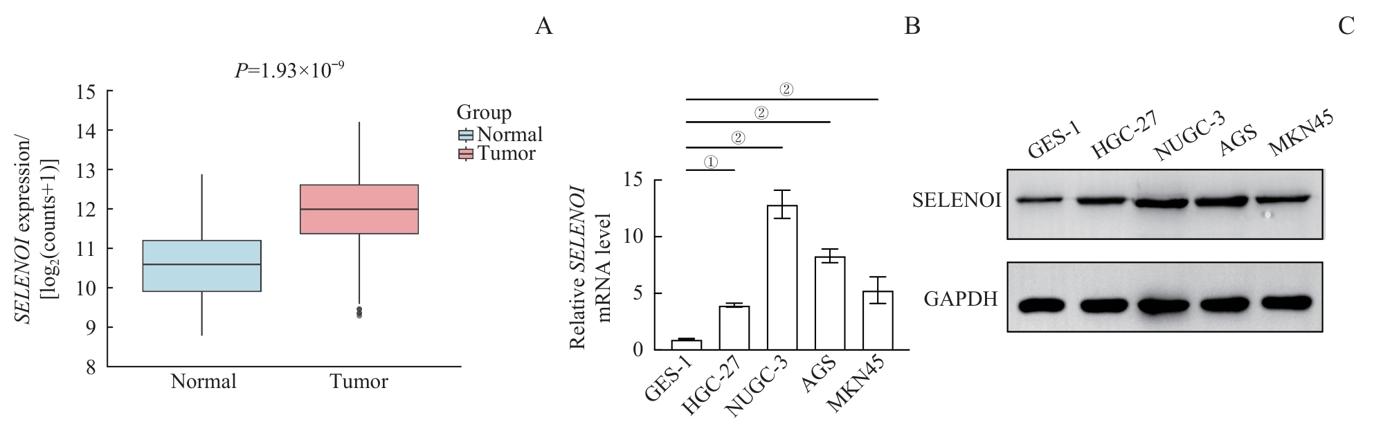
图1 SELENOI在胃癌中的表达水平Note: A. Upregulated expression of SELENOI in tumor tissues compared with normal tissues in the TCGA-STAD cohort. B/C. Elevated SELENOI expression levels in gastric cancer cell lines (HGC-27, NUGC-3, AGS, and MKN45) compared with GES-1 normal gastric epithelial cells, measured by qPCR (B) and Western blotting (C), respectively. ①P=0.004, ②P<0.001.
Fig 1 Expression levels of SELENOI in gastric cancer
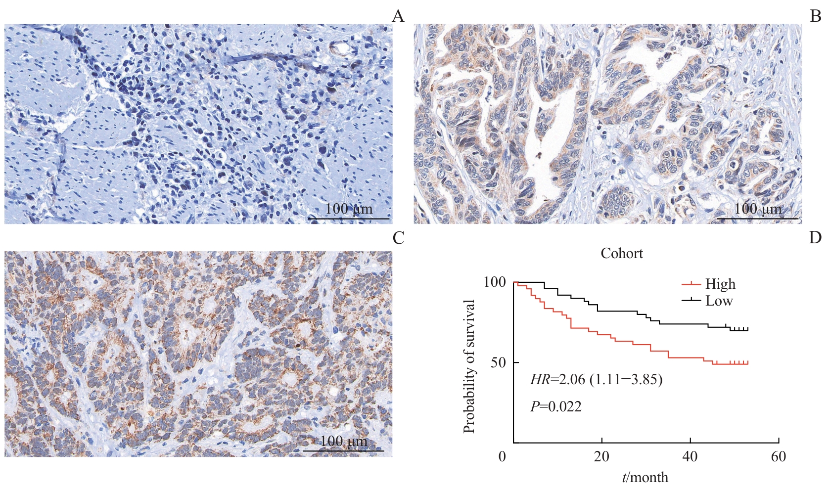
图2 SELENOI表达与STAD临床特征的相关性分析Note: A‒C. Immunohistochemical staining of SELENOI protein in 100 STAD tissue specimens. Representative immunohistochemical images of stage Ⅰ (A, IRS=0), stage Ⅱ (B, IRS=4), and stage Ⅲ (C, IRS=8) STAD tissues. D. Immunohistochemical quantification of SELENOI protein expression in 100 STAD tissues, classification of patients into SELENOI-high and SELENOI-low expression groups, and Kaplan-Meier survival analysis of the association between SELENOI expression and patient survival.
Fig 2 Correlation analysis of SELENOI expression and clinical characteristics of STAD
| Characteristic | SELENOI high-expression group (n=50) | SELENOI low-expression group (n=50) | t/Z/χ² value | P value |
|---|---|---|---|---|
| Age/year | 64.80±10.97 | 62.62±12.34 | 0.934 | 0.386 |
| Gender/n(%) | 0.049 | 0.826 | ||
| Male | 35 (70.0) | 36 (72.0) | ||
| Female | 15 (30.0) | 14 (28.0) | ||
| T stage/n(%) | 0.798 | 0.425 | ||
| T1 | 1 (2.0) | 1 (2.0) | ||
| T2 | 1 (2.0) | 3 (6.0) | ||
| T3 | 6 (12.0) | 7 (14.0) | ||
| T4 | 42 (84.0) | 39 (78.0) | ||
| N stage/n(%) | 0.279 | 0.780 | ||
| N0 | 10 (20.0) | 12 (24.0) | ||
| N1 | 10 (20.0) | 7 (14.0) | ||
| N2 | 12 (24.0) | 15 (30.0) | ||
| N3 | 18 (36.0) | 16 (32.0) | ||
| Clinical stage/n(%) | 0.713 | 0.476 | ||
| Ⅰ | 0 (0) | 1 (2.0) | ||
| Ⅱ | 13 (26.0) | 15 (30.0) | ||
| Ⅲ | 37 (74.0) | 34 (68.0) | ||
| Ⅳ | 0 (0) | 0 (0) |
表3 STAD患者不同SELENOI表达组临床病理特征的比较
Tab 3 Comparison of clinicopathological characteristics between patients with high and low SELENOI expression
| Characteristic | SELENOI high-expression group (n=50) | SELENOI low-expression group (n=50) | t/Z/χ² value | P value |
|---|---|---|---|---|
| Age/year | 64.80±10.97 | 62.62±12.34 | 0.934 | 0.386 |
| Gender/n(%) | 0.049 | 0.826 | ||
| Male | 35 (70.0) | 36 (72.0) | ||
| Female | 15 (30.0) | 14 (28.0) | ||
| T stage/n(%) | 0.798 | 0.425 | ||
| T1 | 1 (2.0) | 1 (2.0) | ||
| T2 | 1 (2.0) | 3 (6.0) | ||
| T3 | 6 (12.0) | 7 (14.0) | ||
| T4 | 42 (84.0) | 39 (78.0) | ||
| N stage/n(%) | 0.279 | 0.780 | ||
| N0 | 10 (20.0) | 12 (24.0) | ||
| N1 | 10 (20.0) | 7 (14.0) | ||
| N2 | 12 (24.0) | 15 (30.0) | ||
| N3 | 18 (36.0) | 16 (32.0) | ||
| Clinical stage/n(%) | 0.713 | 0.476 | ||
| Ⅰ | 0 (0) | 1 (2.0) | ||
| Ⅱ | 13 (26.0) | 15 (30.0) | ||
| Ⅲ | 37 (74.0) | 34 (68.0) | ||
| Ⅳ | 0 (0) | 0 (0) |
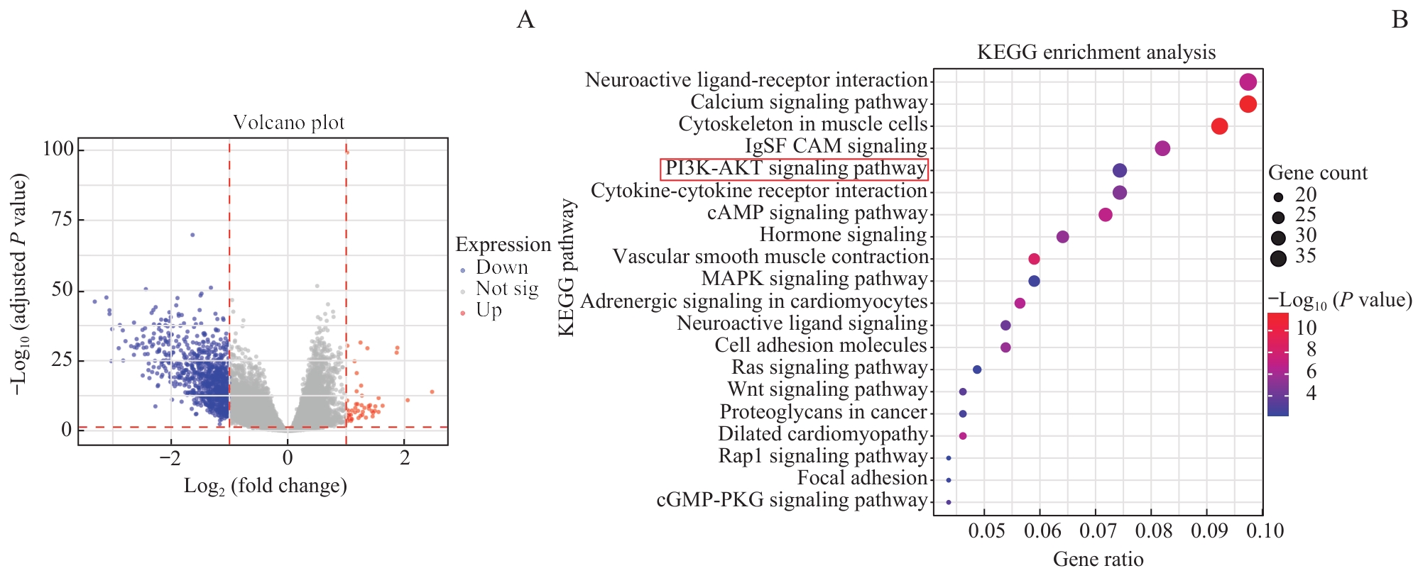
图3 SELENOI 表达相关差异基因的KEGG通路富集分析Note: A. Volcano plot showing differentially expressed genes (TCGA-STAD cohort). B. KEGG pathway enrichment analysis of SELENOI-associated differentially expressed genes (TCGA-STAD cohort).
Fig 3 KEGG pathway enrichment analysis of SELENOI-associated differentially expressed genes in TCGA-STAD
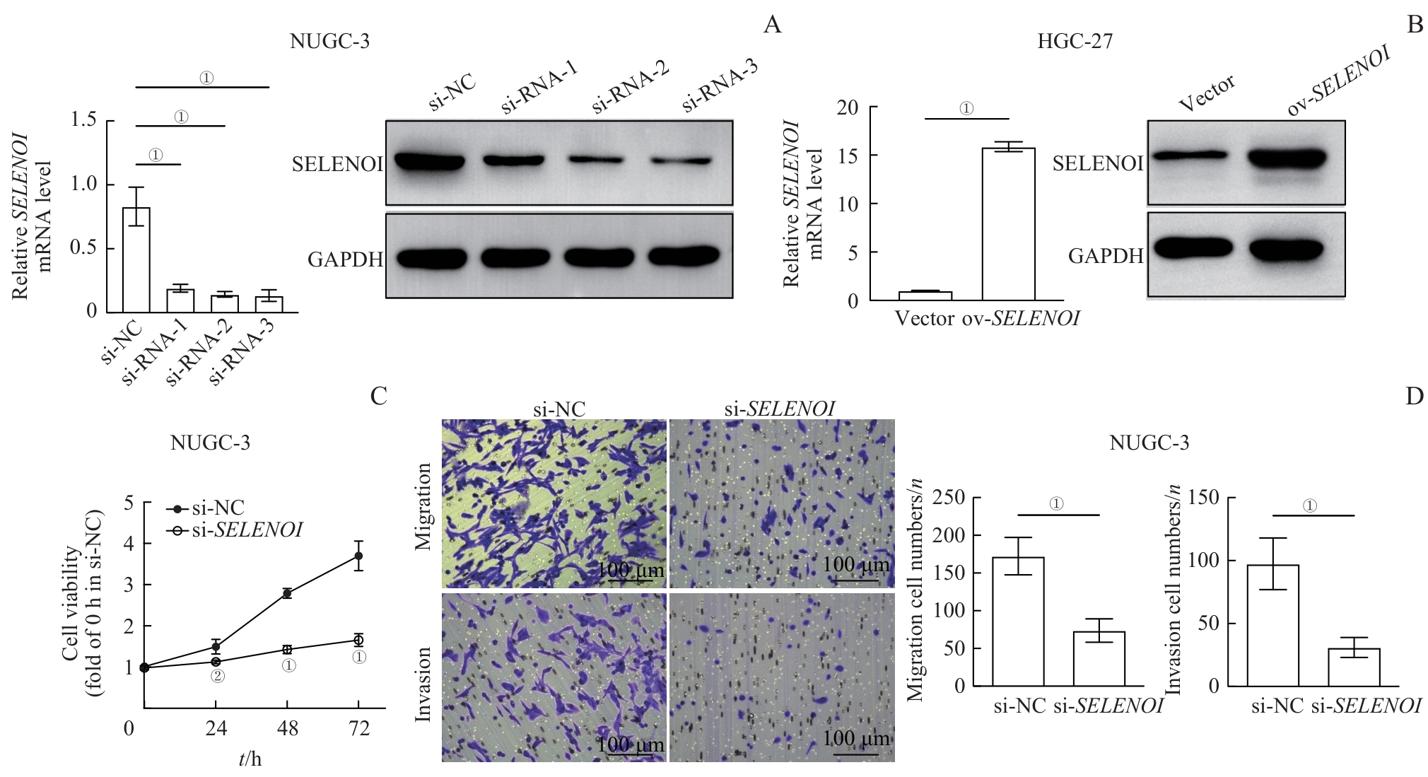
图4 沉默及过表达 SELENOI 对胃癌细胞增殖、迁移及侵袭能力的影响Note: A. Assessment of SELENOI knockdown efficiency after si-SELENOI transfection by qPCR and Western blotting. B. Confirmation of SELENOI overexpression plasmid efficiency by qPCR and Western blotting. C. Impact of si-SELENOI on the proliferation of gastric cancer cell lines by CCK-8 assay. Statistical comparisons were made with the si-NC group. D. Impact of si-SELENOI on the migration and invasion capacities of gastric cancer cell lines determined by Transwell assay. ①P<0.001, ②P=0.025.
Fig 4 Effect of SELENOI silencing and overexpression on the proliferation, migration, and invasion abilities of gastric cancer cells
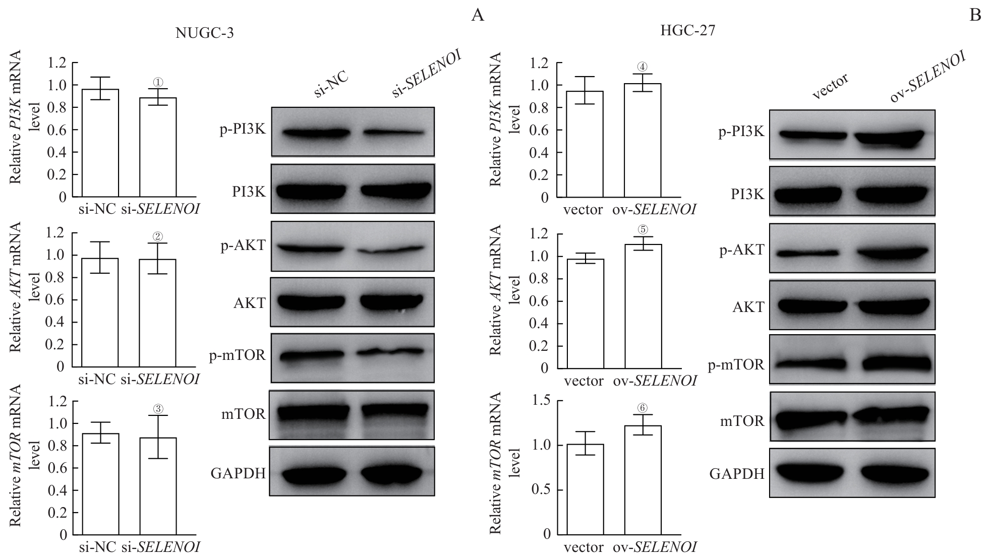
图5 SELENOI对PI3K/AKT/mTOR信号通路的调控Note: A. Effects of si-SELENOI on the expression of the PI3K/AKT/mTOR pathway components detected byqPCR and Western blotting. B. Effects of SELENOI overexpression on the expression of the PI3K/AKT/mTOR pathway components determined by qPCR and Western blotting. ①P=0.349, ②P>0.999, ③P=0.772, compared with the si-NC group; ④P=0.468, ⑤P=0.100, ⑥P=0.106, compared with the vector group.
Fig 5 Regulation of the PI3K/AKT/mTOR signaling pathway by SELENOI
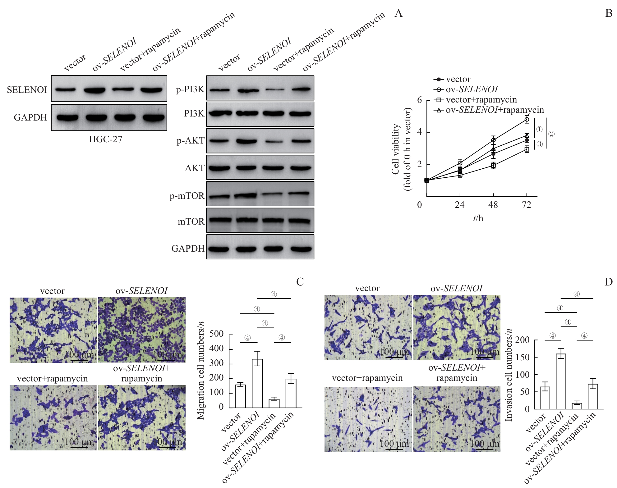
图6 SELENOI通过调控PI3K/AKT/mTOR通路促进胃癌细胞增殖、迁移和侵袭Note: A. Western blotting analysis of PI3K/AKT/mTOR signaling pathway components in SELENOI overexpression HGC-27 gastric cancer cells treated with rapamycin. B. CCK-8 assay assessing the proliferation of HGC-27 gastric cancer cells overexpressing SELENOI and incubated with rapamycin. C/D. Transwell assays examining the migration (C) and invasion (D) capacities of rapamycin-treated HGC-27 gastric cancer cells with SELENOI overexpression. ①P=0.029, ②P=0.015, ③P=0.008, ④P<0.001.
Fig 6 SELENOI promotes the proliferation, migration and invasion of gastric cancer cells through regulation of the PI3K/AKT/mTOR pathway
| [1] | 吴琪, 范伯男, 李岩. 2022全球癌症统计报告分析解读: 中国与世界癌症疾病负担与流行趋势[J]. 诊断学理论与实践, 2025, 24(2): 135-145. |
| Wu Q, Fan B N, Li Y. Analysis and interpretation of the 2022 Global Cancer Statistics Report: cancer burden and epidemiological trends in China and the world[J]. Journal of Diagnostics: Concepts & Practice, 2025, 24(2): 135-145. | |
| [2] | Zheng R S, Zhang S W, Zeng H M, et al. Cancer incidence and mortality in China, 2016[J]. J Natl Cancer Cent, 2022, 2(1): 1-9. |
| [3] | Han B F, Zheng R S, Zeng H M, et al. Cancer incidence and mortality in China, 2022[J]. J Natl Cancer Cent, 2024, 4(1): 47-53. |
| [4] | Henneberry A L, Lagace T A, Ridgway N D, et al. Phosphatidylcholine synthesis influences the diacylglycerol homeostasis required for SEC14p-dependent Golgi function and cell growth[J]. Mol Biol Cell, 2001, 12(3): 511-520. |
| [5] | Nunes L G A, Ma C, Pitts M W, et al. Insights from selenoprotein I mouse models for understanding biological roles of this enzyme[J]. Arch Biochem Biophys, 2025, 768: 110394. |
| [6] | Horibata Y, Maeda S, Konishi A. A Gly-to-Ala substitution confers choline phosphotransferase activity on the CDP-ethanolamine-specific mammalian EPT1[J]. Genes Cells, 2026, 31(2): e70095. |
| [7] | Ma C, Hoffmann F W, Shay A E, et al. Upregulated selenoprotein I during lipopolysaccharide-induced B cell activation promotes lipidomic changes and is required for effective differentiation into IgM-secreting plasma B cells[J]. J Leukoc Biol, 2024, 116(1): 6-17. |
| [8] | Ma C, Hoffmann F W, Marciel M P, et al. Upregulated ethanolamine phospholipid synthesis via selenoprotein I is required for effective metabolic reprogramming during T cell activation[J]. Mol Metab, 2021, 47: 101170. |
| [9] | Ma C, Martinez-Rodriguez V, Hoffmann P R. Roles for selenoprotein I and ethanolamine phospholipid synthesis in T cell activation[J]. Int J Mol Sci, 2021, 22(20): 11174. |
| [10] | Ma C, Hoffmann F W, Nunes L G, et al. Selenoprotein I deficiency in T cells promotes differentiation into tolerant phenotypes while decreasing Th17 pathology[J]. J Leukoc Biol, 2022, 112(6): 1387-1397. |
| [11] | Shi Z, Han Z Y, Chen J Y, et al. Endoplasmic reticulum-resident selenoproteins and their roles in glucose and lipid metabolic disorders[J]. Biochim Biophys Acta Mol Basis Dis, 2024, 1870(6): 167246. |
| [12] | Zhang X, Xiong W, Gao F, et al. Impacts and mechanism of liver-specific knockout of selenoprotein I on hepatic phospholipid metabolism, selenogenome expression, redox status, and resistance to CCl4 toxicity[J]. Free Radic Biol Med, 2025, 235: 426-442. |
| [13] | Huang X, Li T, Yang S H, et al. Hepatocyte-specific SELENOI deficiency predisposes mice to hepatic steatosis and obesity[J]. FASEB J, 2024, 38(11): e23717. |
| [14] | Nunes L G A, Pitts M W, Hoffmann P R. Selenoprotein I (SELENOI) as a critical enzyme in the central nervous system[J]. Arch Biochem Biophys, 2022, 729: 109376. |
| [15] | Nunes L G A, Ma C, Hoffmann F W, et al. Selenoprotein I is indispensable for ether lipid homeostasis and proper myelination[J]. J Biol Chem, 2024, 300(5): 107259. |
| [16] | Huang X, Yang X, Zhang M X, et al. SELENOI functions as a key modulator of ferroptosis pathway in colitis and colorectal cancer[J]. Adv Sci (Weinh), 2024, 11(28): e2404073. |
| [17] | Li J, Chen M M, Huang D W, et al. Inhibition of selenoprotein I promotes ferroptosis and reverses resistance to platinum chemotherapy by impairing Akt phosphorylation in ovarian cancer[J]. MedComm, 2024, 5(12): e70033. |
| [18] | Cao Y J, Luo Y C, Zou J, et al. Autophagy and its role in gastric cancer[J]. Clin Chim Acta, 2019, 489: 10-20. |
| [19] | Wong H, Yau T. Targeted therapy in the management of advanced gastric cancer: are we making progress in the era of personalized medicine?[J]. Oncologist, 2012, 17(3): 346-358. |
| [20] | Byun D S, Cho K, Ryu B K, et al. Frequent monoallelic deletion of PTEN and its reciprocal association with PIK3CA amplification in gastric carcinoma[J]. Int J Cancer, 2003, 104(3): 318-327. |
| [21] | Baghery Saghchy Khorasani A, Pourbagheri-Sigaroodi A, Pirsalehi A, et al. The PI3K/Akt/mTOR signaling pathway in gastric cancer; from oncogenic variations to the possibilities for pharmacologic interventions[J]. Eur J Pharmacol, 2021, 898: 173983. |
| [22] | Wadhwa R, Song S M, Lee J S, et al. Gastric cancer-molecular and clinical dimensions[J]. Nat Rev Clin Oncol, 2013, 10(11): 643-655. |
| [23] | Sukawa Y, Yamamoto H, Nosho K, et al. HER2 expression and PI3K-Akt pathway alterations in gastric cancer[J]. Digestion, 2014, 89(1): 12-17. |
| [1] | 那迪娜·帕尔哈提, 张鹏善, 徐亦天, 陈赟琪, 黄陈. 人去泛素化酶圆柱瘤蛋白截短体质粒的构建及其对胃癌细胞表型的调控研究[J]. 上海交通大学学报(医学版), 2025, 45(9): 1149-1160. |
| [2] | 杨娜, 刘俊丽, 白静, 杨思怡, 韩继明, 张华华. HENMT1通过激活PI3K-AKT-mTOR信号通路促进胃癌的增殖与迁移[J]. 上海交通大学学报(医学版), 2025, 45(6): 717-726. |
| [3] | 陈勇羽, 黄益仁, 陈哲逸, 周冰倩, 陈诗宇, 郑英霞. 丝氨酸蛋白酶抑制因子1在胃癌中的表达及其促进胃癌发展的作用机制[J]. 上海交通大学学报(医学版), 2025, 45(2): 150-160. |
| [4] | 张舒琼, 柯星, 赵兴贺, 陈晓翠, 郑浩东, 陈惠, 沈立松, 杨俊瑶. LINC01123通过结合ENO1促进胃癌的增殖和糖酵解[J]. 上海交通大学学报(医学版), 2025, 45(11): 1443-1457. |
| [5] | 江爽, 俞继卫. m6A去甲基化酶在胃癌发生发展中的作用机制研究进展[J]. 上海交通大学学报(医学版), 2024, 44(2): 271-277. |
| [6] | 冯奕源, 徐忠匀, 尹雅芙, 王辉, 程维维. 二甲双胍改善由C9ORF72肌萎缩侧索硬化/额颞叶痴呆相关多聚甘氨酸-精氨酸诱导的线粒体损伤[J]. 上海交通大学学报(医学版), 2023, 43(7): 839-847. |
| [7] | 陈奕馨, 程丽珍, 林祎嘉, 苗雅. 2型糖尿病脑病小鼠海马中转录因子EB活性与自噬功能的变化[J]. 上海交通大学学报(医学版), 2023, 43(2): 162-170. |
| [8] | 韩婷, 吕纯鑫, 卓萌, 夏青, 刘腾飞, 吴秀奇, 林晓琳, 肖秀英. 进展期胃癌免疫治疗不良反应的相关因素及预后分析[J]. 上海交通大学学报(医学版), 2022, 42(8): 1053-1061. |
| [9] | 陈鸣, 张靖. 血根碱通过上调m6A甲基转移酶14对胃癌细胞增殖和侵袭的抑制作用[J]. 上海交通大学学报(医学版), 2022, 42(2): 135-141. |
| [10] | 陈彬, 崔洪全, 杨懿瑾, 徐海燕, 张玲. 胃癌免疫相关长链非编码RNA预测模型的构建[J]. 上海交通大学学报(医学版), 2022, 42(10): 1394-1403. |
| [11] | 马江磊, 李晓瑶, 赵世富, 杨德君. 胃癌临床分期诊断方法的应用进展[J]. 上海交通大学学报(医学版), 2021, 41(6): 821-825. |
| [12] | 顾琦晟, 张米粒, 曹灿, 李继坤. 基于TCGA数据库分析胃癌可变剪接与肿瘤免疫的关系[J]. 上海交通大学学报(医学版), 2021, 41(4): 448-458. |
| [13] | 岳犇, 王高明, 杨鹿笛, 崔然, 郁丰荣. 胃癌患者预后相关微RNA预测模型的构建及其应用价值探讨[J]. 上海交通大学学报(医学版), 2021, 41(11): 1436-1445. |
| [14] | 易凌荣, 谭波涛, 詹祖雄, 刘媛, 殷樱, 虞乐华. 跑轮运动对小鼠皮质神经元AKT/mTOR通路活性及运动诱发电位的影响[J]. 上海交通大学学报(医学版), 2021, 41(10): 1285-1289. |
| [15] | 张 靖. 川楝素通过下调环状RNA circDLST对胃癌细胞BGC-823的抑制作用[J]. 上海交通大学学报(医学版), 2020, 40(9): 1202-1206. |
| 阅读次数 | ||||||
|
全文 |
|
|||||
|
摘要 |
|
|||||
