Journal of Shanghai Jiao Tong University (Medical Science) ›› 2025, Vol. 45 ›› Issue (4): 415-425.doi: 10.3969/j.issn.1674-8115.2025.04.003
• Basic research • Previous Articles Next Articles
WANG Renjie1, ZHU Chaoyu2, FANG Yunyun2, XIAO Yuanyuan2, WANG Qianqian2, SONG Wenjing2, WEI Li2( )
)
Received:2024-10-30
Accepted:2024-12-20
Online:2025-04-28
Published:2025-04-28
Contact:
WEI Li
E-mail:18930173636@189.cn
Supported by:CLC Number:
WANG Renjie, ZHU Chaoyu, FANG Yunyun, XIAO Yuanyuan, WANG Qianqian, SONG Wenjing, WEI Li. Effects and mechanisms of liraglutide in ameliorating liver fibrosis in NAFLD mice[J]. Journal of Shanghai Jiao Tong University (Medical Science), 2025, 45(4): 415-425.
Add to citation manager EndNote|Ris|BibTeX
URL: https://xuebao.shsmu.edu.cn/EN/10.3969/j.issn.1674-8115.2025.04.003
| Gene | Forward primer (5′→3′) | Reverse primer (5′→3′) |
|---|---|---|
| TGF-β | TGATACGCCTGAGTGGCTGTCT | CACAAGAGCAGTGAGCGCTGAA |
| Col1a | CCTCAGGGTATTGCTGGACAAC | CAGAAGGACCTTGTTTGCCAGG |
| α-SMA | TGCTGACAGAGGCACCACTGAA | CAGTTGTACGTCCAGAGGCATAG |
| Fn | ACAACACCGAGGTGACTGAGAC | GGACACAACGATGCTTCCTGAG |
| Timp1 | TCTTGGTTCCCTGGCGTACTCT | GTGAGTGTCACTCTCCAGTTTGC |
| β-actin | GTGCTATGTTGCTCTAGACTTCG | ATGCCACAGGATTCCATACC |
Tab 1 Primer sequences
| Gene | Forward primer (5′→3′) | Reverse primer (5′→3′) |
|---|---|---|
| TGF-β | TGATACGCCTGAGTGGCTGTCT | CACAAGAGCAGTGAGCGCTGAA |
| Col1a | CCTCAGGGTATTGCTGGACAAC | CAGAAGGACCTTGTTTGCCAGG |
| α-SMA | TGCTGACAGAGGCACCACTGAA | CAGTTGTACGTCCAGAGGCATAG |
| Fn | ACAACACCGAGGTGACTGAGAC | GGACACAACGATGCTTCCTGAG |
| Timp1 | TCTTGGTTCCCTGGCGTACTCT | GTGAGTGTCACTCTCCAGTTTGC |
| β-actin | GTGCTATGTTGCTCTAGACTTCG | ATGCCACAGGATTCCATACC |
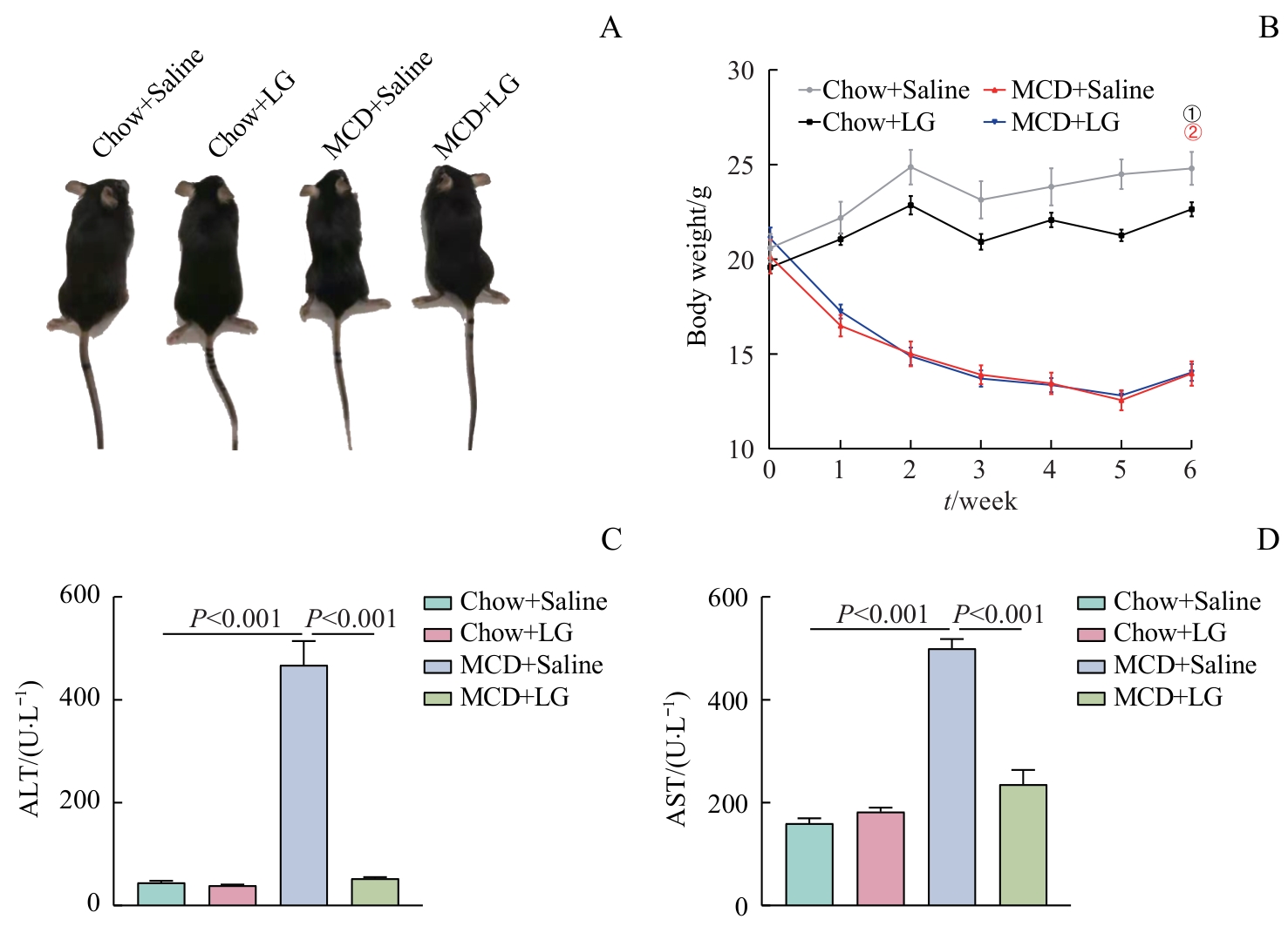
Fig 1 Effect of liraglutide on body weight and liver function in mice
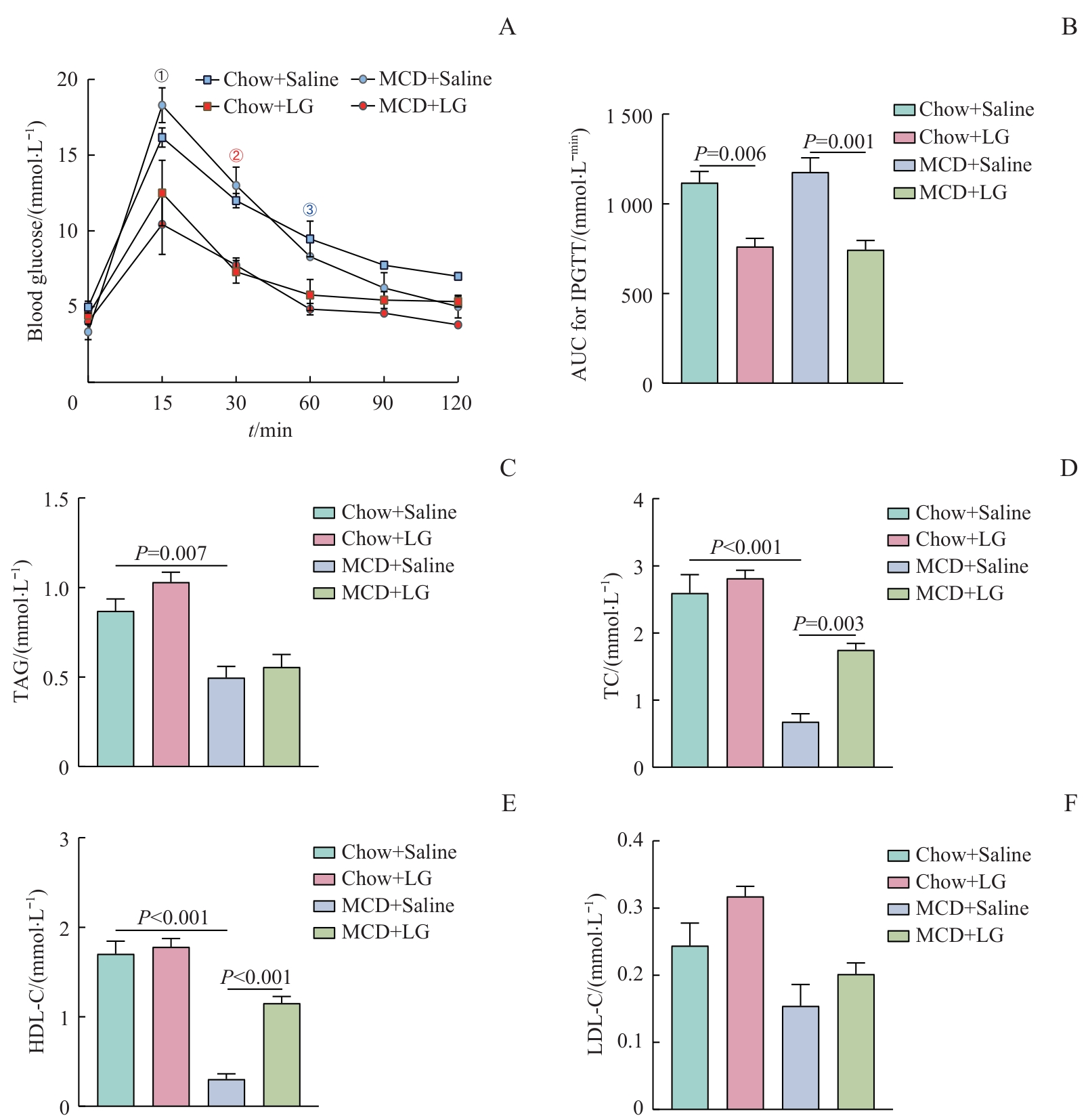
Fig 2 Effects of liraglutide on glucose metabolism and lipid metabolism in mice
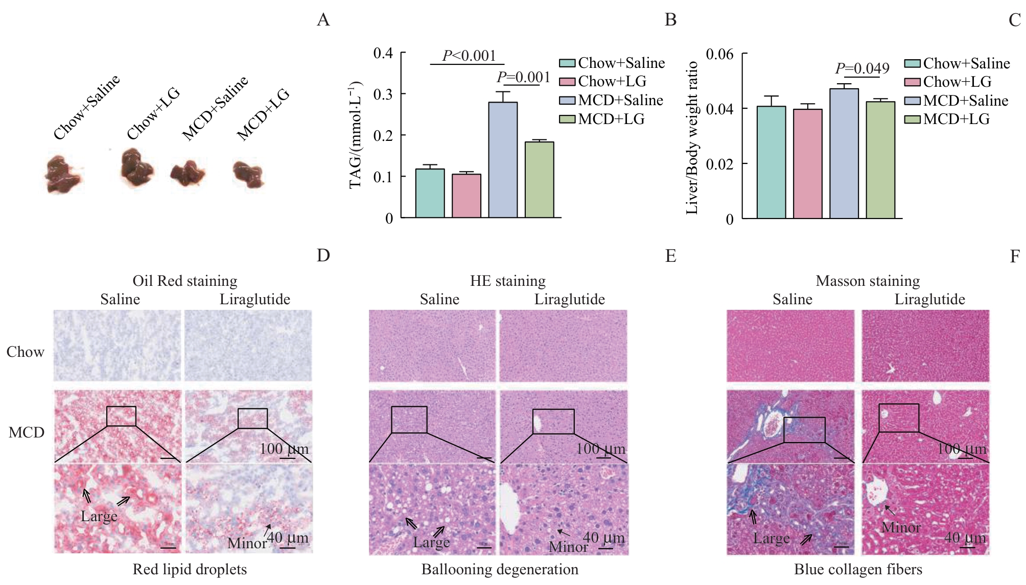
Fig 3 Effect of liraglutide on hepatic steatosis and fibrosis in mice
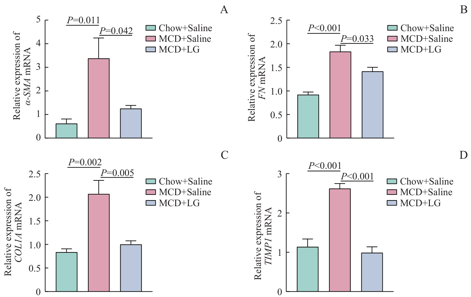
Fig 4 Effect of liraglutide on indices of liver fibrosis genes in mice
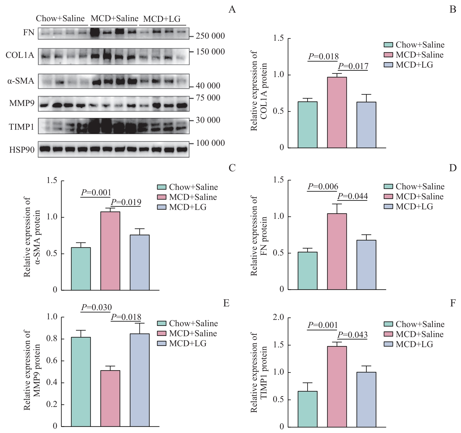
Fig 5 Effect of liraglutide on indices of liver fibrosis protein in mice
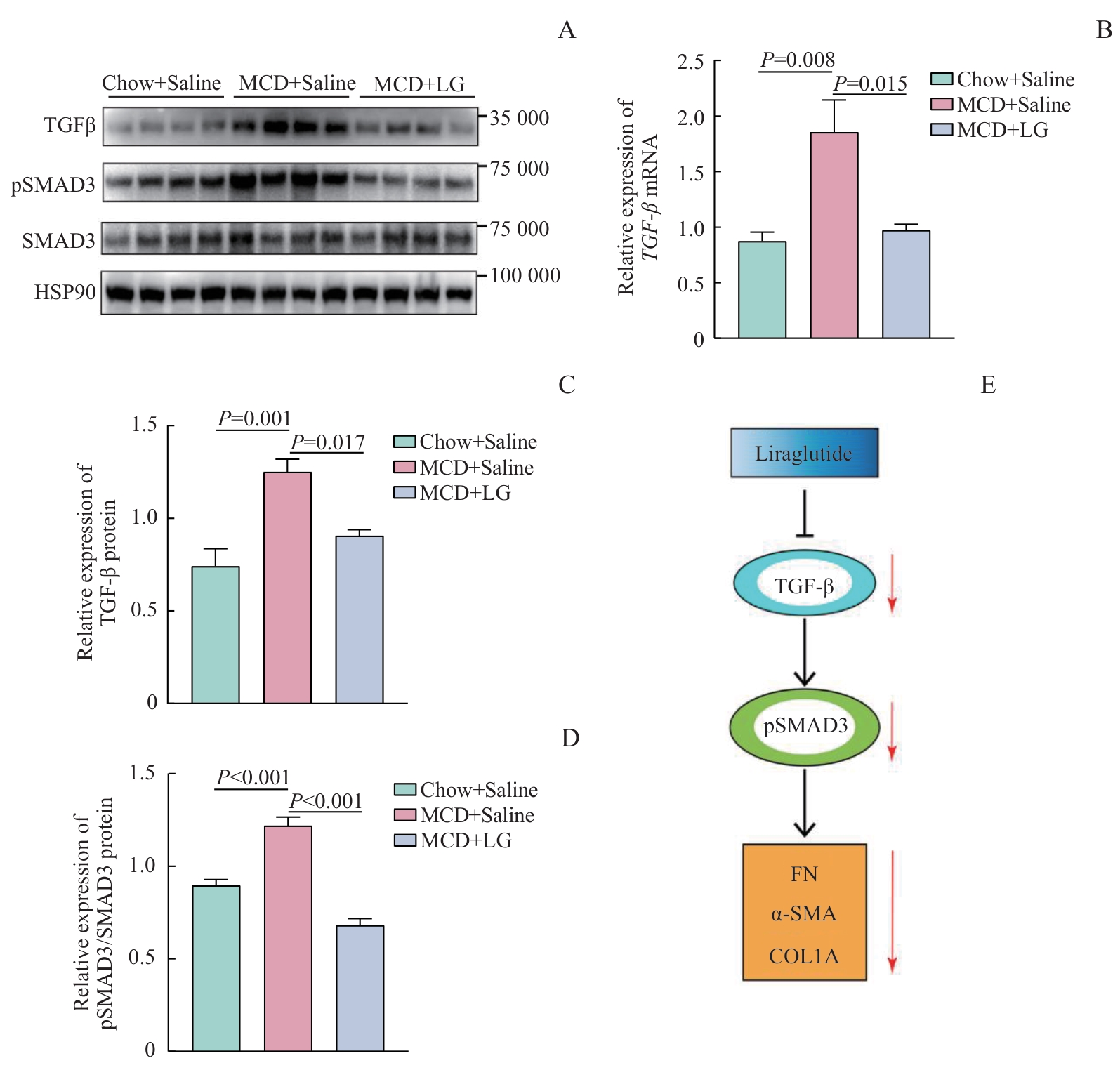
Fig 6 Effect of liraglutide on indices of liver TGF-β pathway in mice
| 1 | 中华医学会肝病学分会脂肪肝和酒精性肝病学组, 中国医师协会脂肪性肝病专家委员会. 非酒精性脂肪性肝病防治指南(2018更新版)[J]. 中华肝脏病杂志, 2018, 26(3): 195-203. |
| National Workshop on Fatty Liver and Alcoholic Liver Disease, Chinese Society of Hepatology, Chinese Medical Association, Fatty Liver Expert Committee, Chinese Medical Doctor Association. Guidelines of prevention and treatment for nonalcoholic fatty liver disease: a 2018 update[J]. Chinese Journal of Hepatology, 2018, 26(3): 195-203. | |
| 2 | JUANOLA O, MARTÍNEZ-LÓPEZ S, FRANCÉS R, et al. Non-alcoholic fatty liver disease: metabolic, genetic, epigenetic and environmental risk factors[J]. Int J Environ Res Public Health, 2021, 18(10): 5227. |
| 3 | DAY C P, JAMES O F W. Steatohepatitis: a tale of two "hits"?[J]. Gastroenterology, 1998, 114(4): 842-845. |
| 4 | WYNN T A, RAMALINGAM T R. Mechanisms of fibrosis: therapeutic translation for fibrotic disease[J]. Nat Med, 2012, 18(7): 1028-1040. |
| 5 | 中华医学会肝病学分会. 代谢相关(非酒精性)脂肪性肝病防治指南(2024年版)[J]. 中华肝脏病杂志, 2024, 32(5): 418-434. |
| Chinese Society of Hepatology, Chinese Medical Association. Guidelines for the prevention and treatment of metabolic dysfunction-associated (non-alcoholic) fatty liver disease (version 2024)[J]. Chinese Journal of Hepatology, 2024, 32(5): 418-434. | |
| 6 | KEAM S J. Resmetirom: first approval[J]. Drugs, 2024, 84(6): 729-735. |
| 7 | SANYAL A J, BEDOSSA P, FRAESSDORF M, et al. A phase 2 randomized trial of survodutide in MASH and fibrosis[J]. N Engl J Med, 2024, 391(4): 311-319. |
| 8 | MÜLLER T D, FINAN B, BLOOM S R, et al. Glucagon-like peptide 1 (GLP-1)[J]. Mol Metab, 2019, 30: 72-130. |
| 9 | 中华医学会内分泌学分会, 中华医学会糖尿病学分会. 胰高糖素样肽-1(GLP-1)受体激动剂用于治疗2型糖尿病的临床专家共识[J]. 中华内科杂志, 2020, 59(11): 836-846. |
| Chinese Society of Endocrinology, Chinese Diabetes Society. Consensus recommendations on utilizing glucagon-like peptide-1 (GLP-1) receptor agonists in the treatment of type 2 diabetes mellitus[J]. Chinese Journal of Internal Medicine, 2020, 59(11): 836-846. | |
| 10 | ARMSTRONG M J, GAUNT P, AITHAL G P, et al. Liraglutide safety and efficacy in patients with non-alcoholic steatohepatitis (LEAN): a multicentre, double-blind, randomised, placebo-controlled phase 2 study[J]. Lancet, 2016, 387(10019): 679-690. |
| 11 | SOMM E, MONTANDON S A, LOIZIDES-MANGOLD U, et al. The GLP-1R agonist liraglutide limits hepatic lipotoxicity and inflammatory response in mice fed a methionine-choline deficient diet[J]. Transl Res, 2021, 227: 75-88. |
| 12 | DELLA PEPA G, PATRÍCIO B G, CARLI F, et al. GLP-1 receptor agonist treatment improves fasting and postprandial lipidomic profiles independently of diabetes and weight loss[J]. Diabetes, 2024, 73(10): 1605-1614. |
| 13 | SHARMA S, MELLS J E, FU P P, et al. GLP-1 analogs reduce hepatocyte steatosis and improve survival by enhancing the unfolded protein response and promoting macroautophagy[J]. PLoS One, 2011, 6(9): e25269. |
| 14 | LI Y K, MA D X, WANG Z M, et al. The glucagon-like peptide-1 (GLP-1) analog liraglutide attenuates renal fibrosis[J]. Pharmacol Res, 2018, 131: 102-111. |
| 15 | 吴佳晋, 钟晨, 李大伟, 等. 甲基转移酶3调控pri-miR-21甲基化修饰在糖尿病肾病肾脏纤维化中的作用[J]. 上海交通大学学报(医学版), 2023, 43(1): 1-7. |
| WU J J, ZHONG C, LI D W, et al. Role of methyltransferase like 3 regulating pri-miR-21 methylation in renal fibrosis of diabetes nephropathy[J]. Journal of Shanghai Jiao Tong University (Medical Science), 2023, 43(1): 1-7. | |
| 16 | 康建华, 李明杰, 栾培培, 等. 西方饮食联合小剂量四氯化碳构建非酒精性脂肪性肝炎小鼠模型研究[J]. 上海交通大学学报(医学版), 2020, 40(5): 590-597. |
| KANG J H, LI M J, LUAN P P, et al. Establishment of non-alcoholic steatohepatitis mouse model induced by Western diet combined with low-dose carbon tetrachloride[J]. Journal of Shanghai Jiao Tong University (Medical Science), 2020, 40(5): 590-597. | |
| 17 | KLEINER D E, BRUNT E M, VAN NATTA M, et al. Design and validation of a histological scoring system for nonalcoholic fatty liver disease[J]. Hepatology, 2005, 41(6): 1313-1321. |
| 18 | PONTES-DA-SILVA R M, DE SOUZA MARINHO T, DE MACEDO CARDOSO L E, et al. Obese mice weight loss role on nonalcoholic fatty liver disease and endoplasmic reticulum stress treated by a GLP-1 receptor agonist[J]. Int J Obes (Lond), 2022, 46(1): 21-29. |
| 19 | ZHOU R, LIN C M, CHENG Y Z, et al. Liraglutide alleviates hepatic steatosis and liver injury in T2MD rats via a GLP-1R dependent AMPK pathway[J]. Front Pharmacol, 2021, 11: 600175. |
| 20 | ZHOU J Y, POUDEL A, WELCHKO R, et al. Liraglutide improves insulin sensitivity in high fat diet induced diabetic mice through multiple pathways[J]. Eur J Pharmacol, 2019, 861: 172594. |
| 21 | BAO Y L, WANG L, PAN H T, et al. Animal and organoid models of liver fibrosis[J]. Front Physiol, 2021, 12: 666138. |
| 22 | 张琪娟, 李继斌, 肖晓秋, 等. 蛋氨酸和胆碱缺乏饮食诱导非酒精性脂肪肝炎的作用及性别差异[J]. 上海交通大学学报(医学版), 2014, 34(1): 30-35, 47. |
| ZHANG Q J, LI J B, XIAO X Q, et al. Gender difference and effects in methionine and choline deficient diets-induced non-alcoholic steatohepatitis[J]. Journal of Shanghai Jiao Tong University (Medical Science), 2014, 34(1): 30-35, 47. | |
| 23 | WU Y R, SHI X Y, MA C Y, et al. Liraglutide improves lipid metabolism by enhancing cholesterol efflux associated with ABCA1 and ERK1/2 pathway[J]. Cardiovasc Diabetol, 2019, 18(1): 146. |
| 24 | 中华医学会肝病学分会, 中华医学会消化病学分会, 中华医学会感染病学分会. 肝纤维化诊断及治疗共识(2019年)[J]. 中华肝脏病杂志, 2019, 27(9): 657-667. |
| Chinese Society of Hepatology Chinese Medical Association, Chinese Society of Gastroenterology Chinese Medical Association, Chinese Society of Infectious Diseases, Chinese Medical Association. Consensus on the diagnosis and therapy of hepatic fibrosis in 2019[J]. Chinese Journal of Hepatology, 2019, 27(9): 657-667. | |
| 25 | HORN P, TACKE F. Metabolic reprogramming in liver fibrosis[J]. Cell Metab, 2024, 36(7): 1439-1455. |
| 26 | ROEHLEN N, CROUCHET E, BAUMERT T F. Liver fibrosis: mechanistic concepts and therapeutic perspectives[J]. Cells, 2020, 9(4): 875. |
| 27 | LIU X Y, LIU R X, HOU F, et al. Fibronectin expression is critical for liver fibrogenesis in vivo and in vitro[J]. Mol Med Rep, 2016, 14(4): 3669-3675. |
| 28 | SOTTILE J, HOCKING D C. Fibronectin polymerization regulates the composition and stability of extracellular matrix fibrils and cell-matrix adhesions[J]. Mol Biol Cell, 2002, 13(10): 3546-3559. |
| 29 | MENG X M, NIKOLIC-PATERSON D J, LAN H Y. TGF-β: the master regulator of fibrosis[J]. Nat Rev Nephrol, 2016, 12(6): 325-338. |
| 30 | TSUCHIDA T, FRIEDMAN S L. Mechanisms of hepatic stellate cell activation[J]. Nat Rev Gastroenterol Hepatol, 2017, 14(7): 397-411. |
| 31 | TRIVEDI P, WANG S, FRIEDMAN S L. The power of plasticity-metabolic regulation of hepatic stellate cells[J]. Cell Metab, 2021, 33(2): 242-257. |
| 32 | ONG C H, THAM C L, HARITH H H, et al. TGF-β-induced fibrosis: a review on the underlying mechanism and potential therapeutic strategies[J]. Eur J Pharmacol, 2021, 911: 174510. |
| 33 | CHILVERY S, BANSOD S, SAIFI M A, et al. Piperlongumine attenuates bile duct ligation-induced liver fibrosis in mice via inhibition of TGF-β1/Smad and EMT pathways[J]. Int Immunopharmacol, 2020, 88: 106909. |
| 34 | WANG Y L, JIAO L K, QIANG C X, et al. The role of matrix metalloproteinase 9 in fibrosis diseases and its molecular mechanisms[J]. Biomed Pharmacother, 2024, 171: 116116. |
| 35 | MURPHY F R, ISSA R, ZHOU X Y, et al. Inhibition of apoptosis of activated hepatic stellate cells by tissue inhibitor of metalloproteinase-1 is mediated via effects on matrix metalloproteinase inhibition: implications for reversibility of liver fibrosis[J]. J Biol Chem, 2002, 277(13): 11069-11076. |
| 36 | ZHANG J L, YANG A H, WU Y, et al. Stachydrine ameliorates carbon tetrachloride-induced hepatic fibrosis by inhibiting inflammation, oxidative stress and regulating MMPs/TIMPs system in rats[J]. Biomed Pharmacother, 2018, 97: 1586-1594. |
| [1] | SONG Jing, JIANG Shuo, WAN Fangyu, LI Juan, MUHETA Adina, MIN Xinying, ZHOU Jingqi. Research progress in effects and mechanisms of dietary pattern interventions in metabolic associated fatty liver disease [J]. Journal of Shanghai Jiao Tong University (Medical Science), 2025, 45(7): 926-933. |
| [2] | BIAN Shu, YU Qian, LIU Liangming. Research progress in the role of endo cannabinoid system in liver diseases [J]. Journal of Shanghai Jiao Tong University (Medical Science), 2024, 44(10): 1299-1306. |
| [3] | WANG Renjie, HUA Hui, ZHU ChaoYu, WEI Li. Advances of GADD45b in hepatic glucose and lipid metabolism [J]. Journal of Shanghai Jiao Tong University (Medical Science), 2024, 44(10): 1316-1322. |
| [4] | BENEDICK Jun Er Chin, SON Peng, ZHANG Yifan, WANG Junqing, GUO Simin. Research progress of the impact of nonalcoholic fatty liver disease on chronic hepatitis B infection [J]. Journal of Shanghai Jiao Tong University (Medical Science), 2023, 43(12): 1585-1590. |
| [5] | LIU Junjun, LU Sumei, ZHANG Bingyang, LI Yongqing, MA Wanshan. Analysis of m6A methylation expression profiles in liver tissue of high-fat diet-induced mouse models of NAFLD [J]. Journal of Shanghai Jiao Tong University (Medical Science), 2023, 43(10): 1227-1235. |
| [6] | Xiaowen ZHANG, Yi WANG, Chan ZHANG, Di ZHANG, Hang YUN, Di HUANG. Effects of Pcsk9 gene interference on high fat-induced nonalcoholic fatty liver disease with atherosclerosis in rats [J]. JOURNAL OF SHANGHAI JIAOTONG UNIVERSITY (MEDICAL SCIENCE), 2022, 42(2): 150-157. |
| [7] | Nan WANG, Ye LU, Feng-jie HAO, Jun-qing WANG. Ameliorative effect of atorvastatin on hepatic fibrosis and its mechanism [J]. JOURNAL OF SHANGHAI JIAOTONG UNIVERSITY (MEDICAL SCIENCE), 2021, 41(12): 1619-1626. |
| [8] | Chao SANG, Dan-dan LIANG, Guo-xiang XIE, Wei JIA, Tian-lu CHEN. Progress in serological noninvasive diagnostic methods for non-alcoholic fatty liver disease [J]. JOURNAL OF SHANGHAI JIAOTONG UNIVERSITY (MEDICAL SCIENCE), 2021, 41(1): 112-117. |
| [9] | GONG Xu-hua1, ZHU Liang2, CHEN Chao3, QIAN Li-jun1. miR-214 promotes the progression of liver fibrosis by activating hepatic stellate cells [J]. JOURNAL OF SHANGHAI JIAOTONG UNIVERSITY (MEDICAL SCIENCE), 2020, 40(6): 776-784. |
| [10] | LI Yue1, 2, SUN Han-xiao2, SHU Jie2, LI Yong-mei1#, SHENG Hui-ming2#. Anti-inflammatory effects of liraglutide on innate lymphoid cells in mice with inflammatory bowel disease [J]. JOURNAL OF SHANGHAI JIAOTONG UNIVERSITY (MEDICAL SCIENCE), 2020, 40(6): 744-751. |
| [11] | GUO Yi-qiong, WU Qiong, WU Ya-ting, GAO Lu-lu, YANG Jian-jun. Effect of Lycium barbarum polysaccharide and aerobic exercise on rats with non-alcoholic fatty liver disease and its mechanism [J]. , 2020, 40(1): 30-. |
| [12] | SANG Chao1, XIE Guo-xiang2, LIANG Dan-dan1, ZHAO Ai-hua1, JIA Wei1, 2, CHEN Tian-lu1. Improvement of liver fibrosis diagnostic models based on Youden index [J]. , 2019, 39(10): 1156-. |
| [13] | ZHU Fan-fan, YANG Li-zhen. Correlation between the levels of serum thyroid hormones and non-alcoholic fatty liver disease in euthyroid type 2 diabetic patients [J]. , 2018, 38(7): 763-. |
| [14] | JIAN Chao-hui, BAO Yu-qian. Research progress of autophagy in non-alcoholic fatty liver disease [J]. , 2018, 38(6): 690-. |
| [15] | SU Zheng-jia, PAN Xiao-qian, CAO Jiu-mei, WU Fang. Effects of liraglutide on glucose induced of miRNA-146b-3p, inflammatory factors and COX-2 in human umbilical vein endothelial cells [J]. , 2018, 38(12): 1420-. |
| Viewed | ||||||
|
Full text |
|
|||||
|
Abstract |
|
|||||