CD10+ neutrophils promote CD8+ T-cell depletion in mucosal malignant melanoma through the SELPG-SELL pathway
HAO Meiling1, MA Yanni1, MA Xuhui1,2, ZHANG Yanjie1, ZENG Hanlin1,3, CHEN Shanshuang,1
1.Shanghai Institute of Precision Medicine, Shanghai Ninth People's Hospital, Shanghai Jiao Tong University School of Medicine, Shanghai 200125, China
2.Department of Oral & Maxillofacial-Head and Neck Oncology, Shanghai Ninth People's Hospital, Shanghai Jiao Tong University School of Medicine, Shanghai, 200011, China
3.Department of Oncology, Shanghai Ninth People's Hospital, Shanghai Jiao Tong University School of Medicine, Shanghai 200011, China
National Natural Science Foundation of China. 82204421. 82072638 Shanghai Central Government - Guided Local Science and Technology Development Fund. YDZX20223100003007
摘要
目的·通过单细胞转录组分析系统解析黏膜恶性黑色素瘤(mucosal malignant melanoma,MM)与皮肤恶性黑色素瘤(cutaneous malignant melanoma,CM)微环境的免疫组分差异,提示MM中CD8+ T细胞耗竭的关键调控机制。方法·收集整合3例初治MM患者手术样本及3例CM单细胞转录组数据,共纳入36 531个细胞进行分析。数据预处理包括批次校正、严格质控(基于线粒体基因比例及基因数筛选)、无监督聚类及细胞类型注释(依据经典标记基因)。进一步采用差异基因分析(Wilcoxon检验)、基因集富集分析[基于基因本体(Gene Ontology,GO)和京都基因与基因组百科全书(Kyoto Encyclopedia of Genes and Genomes,KEGG)数据库]、细胞互作网络分析及癌症基因组图谱(The Cancer Genome Atlas,TCGA)预后关联分析,全面解析MM与CM肿瘤免疫微环境的异质性。结果·与CM相比,MM中CD8+ T细胞的细胞毒性相关基因(GZMB、IFNG等)表达降低(P<0.001),而耗竭标志物(PDCD1、LAG3等)则显著上调(P<0.001),同时耗竭性CD8+ T细胞亚群的比例增加近6倍[(CM:3.38%) vs (MM:19.26%),P<0.001],且终末耗竭评分更高。此外,MM微环境中的中性粒细胞浸润水平显著高于CM[(CM:1.6%) vs (MM:34.1%),P=0.009]。单细胞分析进一步将其划分为5个亚群。其中,CD10+中性粒细胞亚群(占MM中性粒细胞的23.52%)同时高表达促炎分子(S100A8/A9)和免疫抑制分子(MME/CD10、CD55),且该亚群的髓源性抑制细胞(myeloid-derived suppressor cell,MDSC)特征评分显著高于其他亚群。TCGA及MM队列分析进一步证实,该亚群的高表达基因与患者不良预后独立相关(P=0.049),提示该亚群参与肿瘤免疫抑制作用。机制上,CD10+中性粒细胞通过SELPLG-SELL配体-受体对与耗竭性CD8+ T细胞互作,驱动T细胞功能失活。结论·对MM临床样本进行单细胞转录组测序,并系统比较MM与CM的单细胞免疫图谱,提示MM中CD10+中性粒细胞通过SELPLG-SELL通路诱导CD8+ T细胞终末耗竭,解析了MM免疫治疗耐受的分子机制,为靶向中性粒细胞-T细胞互作提供新策略。
关键词:黏膜恶性黑色素瘤
;
中性粒细胞
;
CD8+耗竭T细胞
;
单细胞转录组
Abstract
Objective ·To compare the immune microenvironment of mucosal malignant melanoma (MM) and cutaneous malignant melanoma (CM) using single-cell transcriptome analysis system, and to elucidate the key regulatory mechanism of CD8+ T cell depletion in MM. Methods ·A total of 36 531 cells from three treatment-naïve MM surgical specimens and three CM samples were subjected to single-cell RNA sequencing. Data pre-processing included batch correction, strict quality control (based on mitochondrial gene ratio and gene number screening), unsupervised clustering, and cell-type annotation (using established marker genes). Differential gene analysis (Wilcoxon test), gene set enrichment analysis [based on Gene Ontology (GO) and Kyoto Encyclopedia of Genes and Genomes (KEGG) databases], cell-cell interaction network analysis, and prognostic association analysis using. The Cancer Genome Atlas (TCGA) were used to comprehensively analyse the heterogeneity of MM and CM tumour immune microenvironment. Results ·Compared with CM, the expression of cytotoxicity-related genes (GZMB, IFNG, etc.) in CD8+ T cells in MM was reduced (P<0.001), while the depletion markers (PDCD1, LAG3, etc.) were significantly up-regulated (P<0.001). The proportion of depleted CD8+ T cell subsets increased by nearly 6 times (CM: 3.38% vs MM: 19.26%, P<0.010), and the terminal depletion score was higher. In addition, the level of neutrophil infiltration in the MM microenvironment was significantly higher than that in CM (CM: 1.6% vs MM: 34.1%, P=0.009), and single-cell analysis further divided it into five subgroups. Among them, the CD10+ neutrophil subset (accounting for 23.52% of MM neutrophils) highly expressed pro-inflammatory molecules (S100A8/A9) and immunosuppressive molecules (MME/CD10, CD55), and the characteristic scores of myeloid-derived suppressor cells (MDSCs) in this subset were significantly higher than those of other subgroups. TCGA and MM cohort analyses further confirmed that the highly expressed genes of this subset were independently associated with poor prognosis (P=0.049), suggesting that this subset was involved in tumour immunosuppressive effects. Mechanistically, CD10+ neutrophils interacted with depleted CD8+ T cells through the SELPLG-SELL ligand-receptor pair, driving T-cell inactivation. Conclusion ·Single-cell transcriptome sequencing was performed on MM clinical samples, and the single-cell immunomes of MM and CM were systematically compared, suggesting that CD10+ neutrophils in MM induce CD8+ T-cell terminal depletion through the SELPLG-SELL pathway. The molecular mechanism of MM immunotherapy tolerance was elucidated, providing a new strategy for targeting neutrophil-T cell interaction.
为深入解析差异表达基因的生物学功能,本研究运用clusterProfiler包(v4.14.4)进行系统的功能富集分析。通过整合基因本体(Gene Ontology,GO)和京都基因与基因组百科全书(Kyoto Encyclopedia of Genes and Genomes,KEGG)通路数据库,对差异表达基因集进行功能注释和通路富集分析。显著性阈值设定为P<0.05,以确保结果的可靠性。
1.8 基因集评分分析
本研究构建了多个特征性基因集用于功能评分:① 采用8个细胞毒性相关基因[穿孔素1(perforin 1,PRF1)、干扰素γ(interferon-γ,IFNG)、颗粒溶素(granulysin,GNLY)、NK细胞颗粒蛋白7(natural killer cell granule protein 7,NKG7)、颗粒酶B(granzyme B,GZMB)、颗粒酶A(granzyme A,GZMA)、半胱氨酸蛋白酶抑制剂F(cystatin F,CST7)和肿瘤坏死因子超家族成员10(tumor necrosis factor superfamily member 10,TNFSF10)]评估T细胞毒性。② 选取5个耗竭标志物[细胞毒性T淋巴细胞相关蛋白4(cytotoxic T-lymphocyte- associated protein 4,CTLA-4)、甲型肝炎病毒细胞受体2(hepatitis a virus cellular receptor 2,HAVCR2/TIM-3)、淋巴细胞活化基因3(lymphocyte-activation gene 3,LAG-3)、程序性细胞死亡蛋白1(programmed cell death protein 1,PD-1)和T细胞免疫受体与Ig和ITIM结构域(T cell immunoreceptor with Ig and ITIM domains,TIGIT)]评估T细胞耗竭状态。③ 定义祖细胞耗竭和终末耗竭特征基因集[13]。④ 建立MDSCs特征基因集[23]。⑤ 基于TAN2_CD10+中性粒细胞亚群的高表达基因,构建正常、炎症及MM患者中性粒细胞亚群的特征谱。所有评分均使用UCell包(v2.10.1)进行计算。
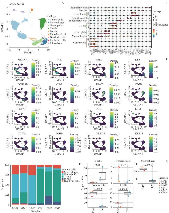
基于单细胞转录组测序技术对3例本院初治MM患者和3例CM患者的样本进行整合分析。对原始测序数据经过严格的质控流程(reads质量评估、低质量细胞过滤等)后,获得了36 531个高质量单细胞数据。采用标准化预处理联合Harmony算法进行批次校正,通过PCA(前20个主成分)及UMAP算法实现降维可视化。基于图论的Leiden无监督聚类(分辨率参数=0.5)将细胞划分为16个转录异质性细胞簇,且2组样本细胞呈现交叉分布特征。通过层级注释策略建立细胞分型体系:首先基于PTPRC(CD45)表达将细胞分为免疫细胞(CD45+,n=16 088)与非免疫细胞(CD45-,n=20 443)二大谱系。非免疫细胞进一步通过经典标记基因鉴定为4类:上皮细胞(KRT14、SPRR3、KRT6A等)、成纤维细胞(COL1A1、COL3A1、DCN等)、内皮细胞(PLVAP、VWF、PECAM1等)及黑色素瘤细胞(DCT、MLANA、EDNRB等)。免疫细胞则细分为五大亚群:B细胞(JCHAIN、IGHG4、MZB1等)、巨噬细胞(LYZ、CD14、C1QC等)、中性粒细胞(FCGR3B、CSF3R、G0S2等)、T细胞(CD3E、TRAC、TRBC2等)及树突状细胞(CD1C、FCER1A、IRF8等)(图1C)。通过UMAP空间映射验证了细胞类型特异性标记的表达模式及分布特征(图1A/B)。细胞类型组成分析显示2种黑色素瘤亚型具有显著的微环境异质性,黏膜亚型表现出独特的免疫特征:T细胞浸润比例较皮肤亚型降低(32.3% vs 7.5%,FDR校正P=0.004),而中性粒细胞比例显著升高(34.1% vs 1.6%,FDR校正P=0.009)(图1D/E)。
Note: A. UMAP map of cell types of mucosal melanosis and cutaneous melanosis. B. Bubble heatmap of the expression of canonical marker genes corresponding to each cell type. C. UMAP map of the expression of the marker genes for the indicated cell subtypes. D/E. Distribution and comparative analysis of immune cells in mucosal melanosis and cutaneous melanosis (MM for mucosal melanosis, CM for cutaneous melanosis) in bar plot (D) and box plot (E).
Fig 1
Single-cell mapping of mucosal malignant melanoma and cutaneous malignant melanoma
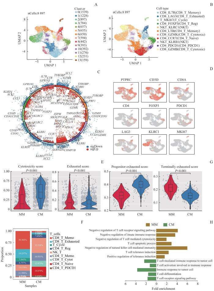
Note: A. UMAP map of T cell clustering in mucosal malignant melanoma and cutaneous malignant melanoma. B. UMAP map of T cell types in mucosal malignant melanoma and cutaneous malignant melanoma. C/D. volcano plot of the highly expressed genes of each T cell population (C) and marker expression maps of T cell types (D) in mucosal malignant melanoma and cutaneous malignant melanoma. E. Cytotoxicity and exhaustion gene set scores of CD8⁺ T cells in mucosal malignant melanoma and cutaneous malignant melanoma. F. Proportional distribution of different types of T cells. G. Progenitor exhaustion and terminal exhaustion gene set scores of exhausted T cells in mucosal malignant melanoma and cutaneous malignant melanoma. H. Differential gene enrichment analysis of exhausted T cells in mucosal malignant melanoma and cutaneous malignant melanoma.
Fig 2
Comparison of T cell characteristics in the immune microenvironment of mucosal malignant melanoma and cutaneous malignant melanoma
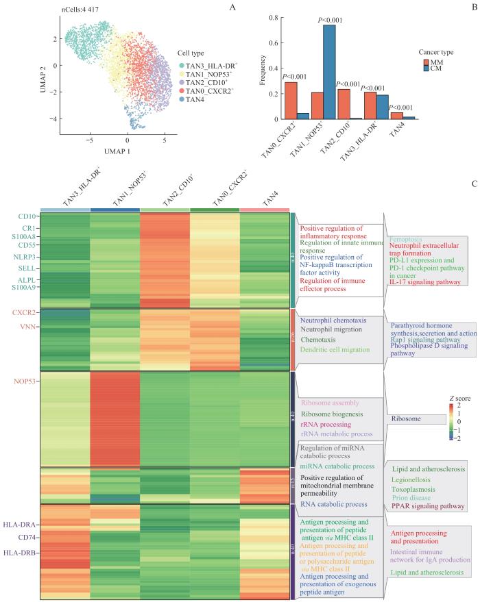
Note: A. UMAP of neutrophil clustering between mucosal and skin melanism. B. Proportion distribution of neutrophil types. C. Analysis of the enrichment of GO and KEGG genes in different subpopulations of neutrophils.
Fig 3
Differences in the characteristics of neutrophil subpopulations in the microenvironment of mucosal and cutaneous malignant melanoma
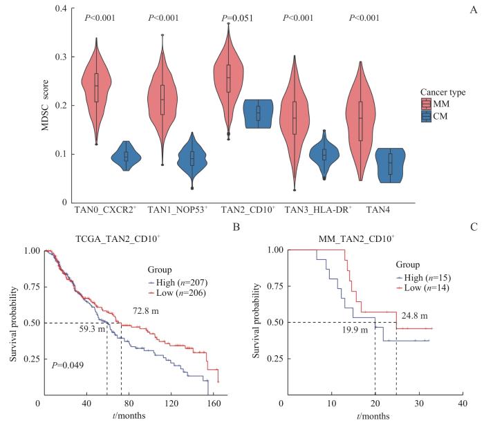
Note: A. MDSC gene set score of neutrophils in mucosal and cutaneous melanosis. B. Prognostic analysis of TAN_CD10+ neutrophil subpopulation infiltration in TCGA-SKCM. C. Prognostic analysis of TAN_CD10+ neutrophil subset infiltration in MM validation set.
Fig 4
Prognostic analysis of TAN2_CD10+ neutrophil subsets in the microenvironment of mucosal and cutaneous malignant melanoma
2.4 MM中TAN2_CD10+ 亚群促进T细胞终末耗竭
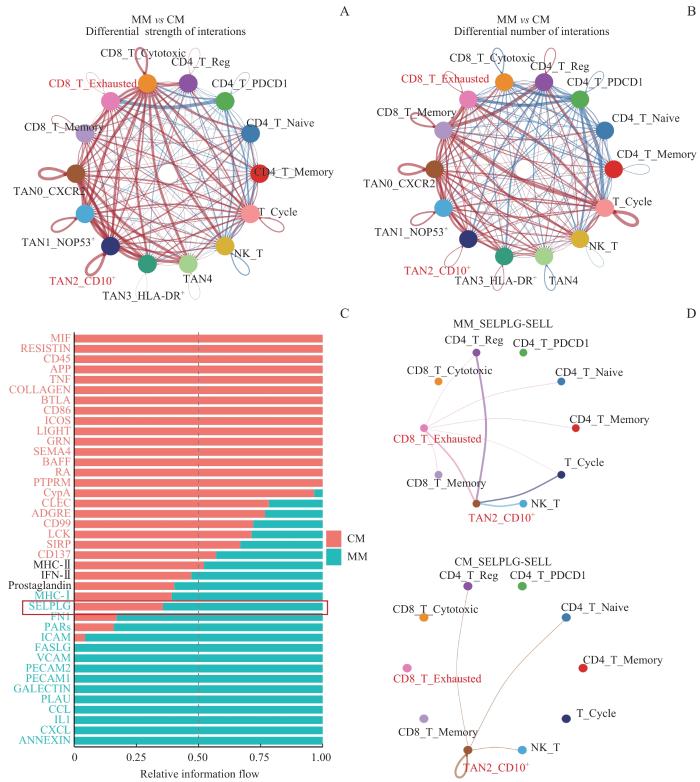
为深入解析TAN2_CD10+亚群与CD8_T_Exhausted之间的免疫调控网络,我们运用Cellchat软件包进行了系统的细胞通信分析。研究结果显示,在MM微环境中,TAN2_CD10+亚群与CD8_T_Exhausted的相互作用呈现出显著增强的特征,其交互数量和信号强度均明显高于CM(图5A/B)。差异信息流网络分析进一步揭示,选择素配体糖蛋白1(selectin ligand glycoprotein 1,SELPLG)信号通路在MM微环境中特异性激活(图5C)。已有研究表明SELPLG-SELL[选择素L(selectin L,SELL)]分子互作能够诱导T细胞耗竭相关的信号转导级联反应,最终导致T细胞功能衰竭[17]。同时,临床前实验证实,靶向阻断SELPLG信号通路可协同增强PD-1/CTLA-4[细胞毒性T淋巴细胞相关蛋白4(cytotoxic t-lymphocyte-associated protein 4,CTLA-4)]抑制剂的治疗效果[37],有效逆转T细胞耗竭状态。通过对比分析不同组织来源的样本,我们发现MM中TAN2_ CD10+亚群通过SELPLG-SELL轴与CD8_T_Exhausted建立的相互作用网络显著强于皮肤组织(图5D)。这些数据表明TAN2_CD10+亚群可能通过SELPLG信号通路抑制T细胞的抗肿瘤功能,提示靶向干预SELPLG信号通路可能成为增强黑色素瘤免疫治疗效果的新策略。
Note: A/B. The differences in the intensity of cellular interactions (A) and the number of interactions (B) in mucosal and skin mellanism are shown in red for upreluation and in blue for downregulation. C. Percentage stacked histogram of the overall information flow of each signaling pathway in mucosal and skin dark. D SELPLG-SELL-mediated cellular interaction network in mucosal and cutaneous malignant melanoma.
Fig 5
Characterization of T cell-neutrophil interaction networks in the microenvironment of mucosal and cutaneous malignant melanoma
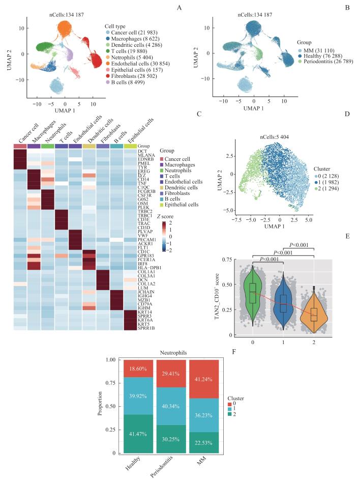
Note: A. UMAP map of cell types in mucosal health, mucosal inflammation, and mucosal melanoma. B. UMAP map of sample distribution in mucosal health, mucosal inflammation, and mucosal melanoma. C. Gene expression spectrum of cell types in mucosal health, mucosal inflammation, and mucosal melanoma. D. UMAP map of neutrophil distribution groups in mucosal health, mucosal inflammation, and mucosal melanoma. E. MDSC gene set score of neutrophil subgroups in mucosal health and mucosal inflammation. F. Cell proportion of neutrophil subgroups in mucosal health and mucosal inflammation.
Fig 6
TAN2_CD10+ neutrophil subpopulation is enriched by inflammation in the microenvironment of mucosal and cutaneous malignant melanoma
HAO Meiling was involved in the data analysis and paper writing. MA Yanni and ZENG Hanlin were responsible for the guidance of the experimental protocol and the analysis of the experimental results. MA Xuhui and ZHANG Yanjie were involved in the collection of the patient samples and clinical information; ZENG Hanlin and CHEN Shanshuang were responsible for the review of the paper. All authors read and agreed to submit the final manuscript.
This research protocol has been reviewed and approved by the Ethics Committee of Shanghai Ninth People′s Hospital, Shanghai Jiao Tong University School of Medicine (No. SH9H-2019-T279-3).
SALEH M, JAVADI S, ELSHERIF S, et al. Multimodality imaging and genetics of primary mucosal melanomas and response to treatment[J]. Radiographics, 2021, 41(7): 1954-1972.
OTTAVIANO M, GIUNTA E F, MARANDINO L, et al. Anorectal and genital mucosal melanoma: diagnostic challenges, current knowledge and therapeutic opportunities of rare melanomas[J]. Biomedicines, 2022, 10(1): 150.
KOTTSCHADE L A, POND G R, OLSZANSKI A J, et al. SALVO: single-arm trial of ipilimumab and nivolumab as adjuvant therapy for resected mucosal melanoma[J]. Clin Cancer Res, 2023, 29(12): 2220-2225.
SI L, FANG M Y, CHEN Y, et al. Atezolizumab in combination with bevacizumab in patients with unresectable locally advanced or metastatic mucosal melanoma: interim analysis of an open-label phase II trial[J]. J Clin Oncol, 2021, 39(15_suppl): 9511.
JOHNSON D B, SWETTER S M, SALAMA A K S, et al. Cutaneous melanoma: management of melanoma brain metastases and molecular testing[J]. J Natl Compr Cancer Netw, 19(5.5): 644-647.
LI S M, WU X W, YAN X Q, et al. Toripalimab plus axitinib in patients with metastatic mucosal melanoma: 3-year survival update and biomarker analysis[J]. J Immunother Cancer, 2022, 10(2): e004036.
BLACKBURN S D, SHIN H, FREEMAN G J, et al. Selective expansion of a subset of exhausted CD8 T cells by alphaPD-L1 blockade[J]. Proc Natl Acad Sci USA, 2008, 105(39): 15016-15021.
ZENG S S, HU H L, LI Z Y, et al. Local TSH/TSHR signaling promotes CD8+ T cell exhaustion and immune evasion in colorectal carcinoma[J]. Cancer Commun (Lond), 2024, 44(11): 1287-1310.
GAO Y, OUYANG Z J, YANG C, et al. Overcoming T cell exhaustion via immune checkpoint modulation with a dendrimer-based hybrid nano complex[J]. Adv Healthc Mater, 2021, 10(19): e2100833.
MILLER B C, SEN D R, AL ABOSY R, et al. Subsets of exhausted CD8+ T cells differentially mediate tumor control and respond to checkpoint blockade[J]. Nat Immunol, 2019, 20(3): 326-336.
KALTENMEIER C, YAZDANI H O, MORDER K, et al. Neutrophil extracellular traps promote T cell exhaustion in the tumor microenvironment[J]. Front Immunol, 2021, 12: 785222.
WANG L W, LIU Y H, DAI Y T, et al. Single-cell RNA-seq analysis reveals BHLHE40-driven pro-tumour neutrophils with hyperactivated glycolysis in pancreatic tumour microenvironment[J]. Gut, 2023, 72(5): 958-971.
MENG Y, YE F, NIE P P, et al. Immunosuppressive CD10+ALPL+ neutrophils promote resistance to anti-PD-1 therapy in HCC by mediating irreversible exhaustion of T cells[J]. J Hepatol, 2023, 79(6): 1435-1449.
HUANG C T, LIAO Y H, LIAU J Y, et al. Pretreatment neutrophil-to-lymphocyte ratio predicts survival in primary mucosal melanoma[J]. J Am Acad Dermatol, 2025, 92(2): 340-343.
SADE-FELDMAN M, YIZHAK K, BJORGAARD S L, et al. Defining T cell states associated with response to checkpoint immunotherapy in melanoma[J]. Cell, 2018, 175(4): 998-1013.e20.
TIROSH I, IZAR B, PRAKADAN S M, et al. Dissecting the multicellular ecosystem of metastatic melanoma by single-cell RNA-seq[J]. Science, 2016, 352(6282): 189-196.
ZHANG C, SHEN H R, YANG T L, et al. A single-cell analysis reveals tumor heterogeneity and immune environment of acral melanoma[J]. Nat Commun, 2022, 13(1): 7250.
ZHOU Z Y, ZHOU X X, JIANG X, et al. Single-cell profiling identifies IL1Bhi macrophages associated with inflammation in PD-1 inhibitor-induced inflammatory arthritis[J]. Nat Commun, 2024, 15(1): 2107.
ALSHETAIWI H, PERVOLARAKIS N, MCINTYRE L L, et al. Defining the emergence of myeloid-derived suppressor cells in breast cancer using single-cell transcriptomics[J]. Sci Immunol, 2020, 5(44): eaay6017.
LIU W, STACHURA P, XU H C, et al. BAFF attenuates immunosuppressive monocytes in the melanoma tumor microenvironment[J]. Cancer Res, 2022, 82(2): 264-277.
LIU X B, WANG Y Y, BAUER A T, et al. Neutrophils activated by membrane attack complexes increase the permeability of melanoma blood vessels[J]. Proc Natl Acad Sci USA, 2022, 119(33): e2122716119.
BARON M, IDEKER T. Abstract 178: desmosome mutations in melanoma promote cellular proliferation and disease progression[J]. Cancer Res, 2021, 81(13_Supplement): 178.
LIU S J, YUAN S J, LIU M C, et al. In situ tumor cell engineering reverses immune escape to enhance immunotherapy effect[J]. Acta Pharm Sin B, 2025, 15(1): 627-641.
SÁNCHEZ-MAGRANER L, MILES J, BAKER C L, et al. High PD-1/PD-L1 checkpoint interaction infers tumor selection and therapeutic sensitivity to anti-PD-1/PD-L1 treatment[J]. Cancer Res, 2020, 80(19): 4244-4257.
MA H Y, YAMAMOTO G, XU J, et al. IL-17 signaling in steatotic hepatocytes and macrophages promotes hepatocellular carcinoma in alcohol-related liver disease[J]. J Hepatol, 2020, 72(5): 946-959.
YANG L Y, LUO Q, LU L, et al. Increased neutrophil extracellular traps promote metastasis potential of hepatocellular carcinoma via provoking tumorous inflammatory response[J]. J Hematol Oncol, 2020, 13(1): 3.
VIRAMONTES K M, NEUBERT E N, DEROGATIS J M, et al. PD-1 immune checkpoint blockade and PSGL-1 inhibition synergize to reinvigorate exhausted T cells[J]. Front Immunol, 2022, 13: 869768.
ZENG W Y, WANG Y, ZHANG Q, et al. Neutrophil nanodecoys inhibit tumor metastasis by blocking the interaction between tumor cells and neutrophils[J]. ACS Nano, 2024, 18(10): 7363-7378.
QUE H Y, FU Q M, LAN T X, et al. Tumor-associated neutrophils and neutrophil-targeted cancer therapies[J]. Biochim Biophys Acta Rev Cancer, 2022, 1877(5): 188762.