
上海交通大学学报(医学版) ›› 2026, Vol. 46 ›› Issue (7): 829-838.doi: 10.3969/j.issn.1674-8115.2026.07.001
• 前沿述评 • 下一篇
沈靖婷, 柳稚旭, 王旭东( )
)
收稿日期:2026-02-28
接受日期:2026-04-29
出版日期:2026-07-28
发布日期:2026-07-28
通讯作者:
王旭东,主任医师,教授,博士;电子信箱:xudongwang70@hotmail.com。基金资助:
Shen Jingting, Liu Zhixu, Wang Xudong()
Received:2026-02-28
Accepted:2026-04-29
Online:2026-07-28
Published:2026-07-28
Contact:
Wang Xudong, E-mail: xudongwang70@hotmail.com.Supported by:摘要:
口腔遗传病与罕见病具有发病率低、表型异质性强且致病机制复杂的特点,病情严重,诊断困难且治疗手段有限。随着多组学测序以及医学影像技术的快速发展,海量的分子数据和临床数据为该类疾病的研究提供了新的视角。人工智能(artificial intelligence,AI)技术凭借其强大的模式识别和复杂关系建模能力,已在口腔遗传病与罕见病基础研究和临床诊疗领域取得显著成果。在致病机制研究方面,AI可解读基因组、转录组、蛋白组及微生物组等多组学数据,识别新的分子标志物并构建疾病预测模型,推动复杂病因的解析。在临床诊断方面,AI显著提升了口腔影像的自动化分析能力,智能诊断龋齿、牙发育异常、唾液腺疾病、骨纤维异常增殖症及多种颅颌面畸形疾病,也为手术治疗规划提供决策支持。尽管AI在口腔遗传病领域取得显著进展,但仍面临数据稀缺、模型可解释性不足及伦理规范限制等挑战。该文综述AI在口腔遗传病与罕见病领域应用的最新进展,系统梳理其在深化致病机制研究和加速临床诊断方面的进展、挑战与前景,旨在推动口腔遗传病精准医学的整体进步及优质医疗资源下沉。
中图分类号:
沈靖婷, 柳稚旭, 王旭东. AI在口腔遗传病与罕见病机制研究及临床诊断中的应用进展[J]. 上海交通大学学报(医学版), 2026, 46(7): 829-838.
Shen Jingting, Liu Zhixu, Wang Xudong. Applications and progress of AI in mechanistic research and clinical diagnosis of oral genetic and rare diseases[J]. Journal of Shanghai Jiao Tong University (Medical Science), 2026,(7): 829-838.
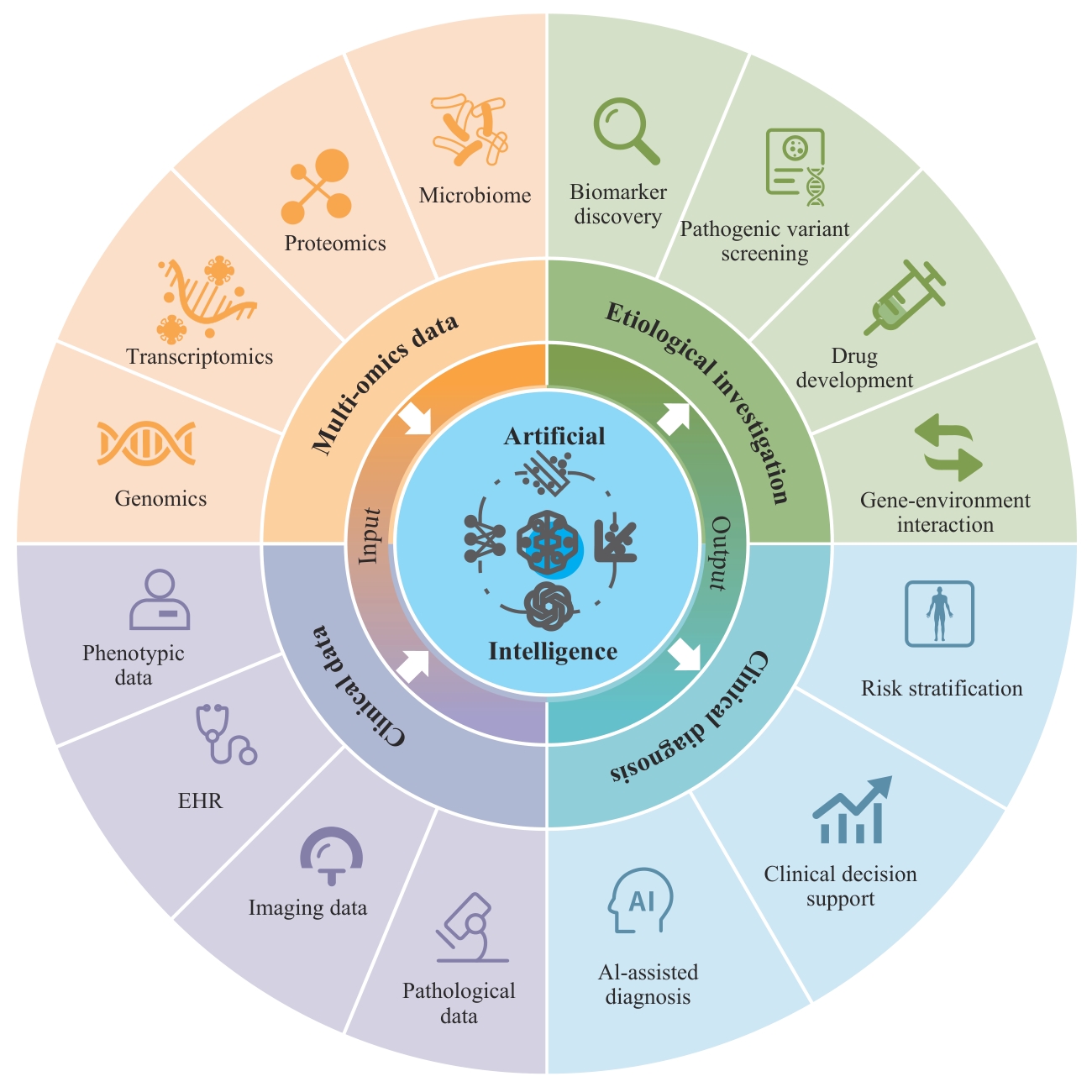
图1 AI在口腔遗传病和罕见病领域中的应用方向
Fig 1 Applications of AI in oral genetic and rare diseasesNote: EHR—electronic health record.
| Reference | Year | Dataset | Objective | AI model |
|---|---|---|---|---|
| Zhang, et al[ | 2018 | 43 GWAS-significant SNPs associated with OFCs | Genetic risk assessment of OFCs | SVM, LR, NB, RF, KNN, DT, and DNN |
| Dai, et al[ | 2024 | Epigenetic sequencing data from PCW4-10 | Pathogenic variant prioritization for OFCs | CNN |
| Forrest, et al[ | 2025 | Training cohort of 1.3 million individuals with 10 genetic diseases | Genetic variant penetrance estimation | XGBoost |
| Kamitaki, et al[ | 2026 | Genomic and oral microbiome data from 12 519 individuals | Genetic variant-oral microbiome association analysis | PCA |
| Guo, et al[ | 2025 | scRNA-seq data of mouse molars from embryonic stages to postnatal days | Cell subtype identification | PCA, UMAP, etc. |
| Huang, et al[ | 2025 | scRNA-seq data of mouse palate during embryonic development | Cell subtype identification | PCA, UMAP, etc. |
| Yankee, et al[ | 2023 | scRNA-seq data of human embryonic craniofacial tissues | Cell subtype identification | PCA, UMAP, etc. |
| Liu, et al[ | 2025 | Paired bulk transcriptomic and single-cell transcriptomic data | Single-cell proteome prediction | Transformer |
表1 AI在致病机制研究中的应用
Tab 1 AI applications in pathogenic mechanism study
| Reference | Year | Dataset | Objective | AI model |
|---|---|---|---|---|
| Zhang, et al[ | 2018 | 43 GWAS-significant SNPs associated with OFCs | Genetic risk assessment of OFCs | SVM, LR, NB, RF, KNN, DT, and DNN |
| Dai, et al[ | 2024 | Epigenetic sequencing data from PCW4-10 | Pathogenic variant prioritization for OFCs | CNN |
| Forrest, et al[ | 2025 | Training cohort of 1.3 million individuals with 10 genetic diseases | Genetic variant penetrance estimation | XGBoost |
| Kamitaki, et al[ | 2026 | Genomic and oral microbiome data from 12 519 individuals | Genetic variant-oral microbiome association analysis | PCA |
| Guo, et al[ | 2025 | scRNA-seq data of mouse molars from embryonic stages to postnatal days | Cell subtype identification | PCA, UMAP, etc. |
| Huang, et al[ | 2025 | scRNA-seq data of mouse palate during embryonic development | Cell subtype identification | PCA, UMAP, etc. |
| Yankee, et al[ | 2023 | scRNA-seq data of human embryonic craniofacial tissues | Cell subtype identification | PCA, UMAP, etc. |
| Liu, et al[ | 2025 | Paired bulk transcriptomic and single-cell transcriptomic data | Single-cell proteome prediction | Transformer |
| Reference | Year | Dataset | Objective | AI model |
|---|---|---|---|---|
| Lee, et al[ | 2018 | 3 000 periapical radiographs | Dental caries detection | CNN |
| Hiraiwa, et al[ | 2019 | CBCT images and panoramic radiographs of mandibular first molars from 400 patients | Root canal morphology classification | CNN |
| Ekert, et al[ | 2019 | 2 001 panoramic radiographs | Periapical lesion diagnosis | DNN |
| Duman, et al[ | 2023 | 220 panoramic radiographs of children aged 6‒9 years | Hyperdontia diagnosis | CNN |
| Incerti Parenti, et al[ | 2025 | A retrospective dataset of 250 panoramic radiographs from patients aged 6‒13 years | Primary and permanent tooth detection and segmentation | CNN |
| Sheng, et al[ | 2023 | 100 panoramic radiographs | Tooth segmentation | Transformer |
| Han, et al[ | 2023 | 203 salivary gland pathology slides | Segmentation of lymphocytic infiltration areas | DNN |
| Chiesa-Estomba, et al[ | 2021 | Multi-source clinical data from 345 patients undergoing salivary gland tumor surgery | Prediction of postoperative facial nerve palsy | KNN, RF, BC, and LDC |
| Jurek, et al[ | 2020 | Fetal ultrasound images | Fetal cleft palate diagnosis | NLP |
| Shafi, et al[ | 2020 | 1 000 prenatal survey records | Risk prediction of cleft lip and palate | DNN |
| Kuwada, et al[ | 2021 | Panoramic radiographs of 383 patients with cleft conditions (with or without alveolar cleft) and 210 healthy controls | Cleft palate classification | DNN |
| Zhang, et al[ | 2020 | CBCT images of 21 patients with unilateral or bilateral CLP | Alveolar bone defect volume estimation | CNN |
| Pactas, et al[ | 2019 | Pre- and post-treatment photographs of 146 orthognathic patients | Assessment of orthognathic surgical outcome | CNN |
| Huang, et al[ | 2025 | Pre- and post-treatment CBCT images of 100 orthognathic surgery patients | Prediction of postoperative facial appearance | CNN |
| Adel, et al[ | 2025 | 3D facial images of 130 patients | Facial symmetry assessment | DNN |
| Chen, et al[ | 2025 | CT images of 15 patients | Soft tissue defect assessment | CNN |
| Saranya, et al[ | 2022 | Bone CT images | FD lesion segmentation | R-CNN |
| Li, et al[ | 2025 | CT images of 148 FD patients | Prediction of FD lesion growth status | CNN |
| Zhang, et al[ | 2024 | CT images of 220 patients with FD or OF | Differential diagnosis between FD and OF | RF, SVM, LightGBM, and XGBoost |
表2 AI在临床诊断和评估中的应用
Tab 2 AI applications in clinical diagnosis and evaluation
| Reference | Year | Dataset | Objective | AI model |
|---|---|---|---|---|
| Lee, et al[ | 2018 | 3 000 periapical radiographs | Dental caries detection | CNN |
| Hiraiwa, et al[ | 2019 | CBCT images and panoramic radiographs of mandibular first molars from 400 patients | Root canal morphology classification | CNN |
| Ekert, et al[ | 2019 | 2 001 panoramic radiographs | Periapical lesion diagnosis | DNN |
| Duman, et al[ | 2023 | 220 panoramic radiographs of children aged 6‒9 years | Hyperdontia diagnosis | CNN |
| Incerti Parenti, et al[ | 2025 | A retrospective dataset of 250 panoramic radiographs from patients aged 6‒13 years | Primary and permanent tooth detection and segmentation | CNN |
| Sheng, et al[ | 2023 | 100 panoramic radiographs | Tooth segmentation | Transformer |
| Han, et al[ | 2023 | 203 salivary gland pathology slides | Segmentation of lymphocytic infiltration areas | DNN |
| Chiesa-Estomba, et al[ | 2021 | Multi-source clinical data from 345 patients undergoing salivary gland tumor surgery | Prediction of postoperative facial nerve palsy | KNN, RF, BC, and LDC |
| Jurek, et al[ | 2020 | Fetal ultrasound images | Fetal cleft palate diagnosis | NLP |
| Shafi, et al[ | 2020 | 1 000 prenatal survey records | Risk prediction of cleft lip and palate | DNN |
| Kuwada, et al[ | 2021 | Panoramic radiographs of 383 patients with cleft conditions (with or without alveolar cleft) and 210 healthy controls | Cleft palate classification | DNN |
| Zhang, et al[ | 2020 | CBCT images of 21 patients with unilateral or bilateral CLP | Alveolar bone defect volume estimation | CNN |
| Pactas, et al[ | 2019 | Pre- and post-treatment photographs of 146 orthognathic patients | Assessment of orthognathic surgical outcome | CNN |
| Huang, et al[ | 2025 | Pre- and post-treatment CBCT images of 100 orthognathic surgery patients | Prediction of postoperative facial appearance | CNN |
| Adel, et al[ | 2025 | 3D facial images of 130 patients | Facial symmetry assessment | DNN |
| Chen, et al[ | 2025 | CT images of 15 patients | Soft tissue defect assessment | CNN |
| Saranya, et al[ | 2022 | Bone CT images | FD lesion segmentation | R-CNN |
| Li, et al[ | 2025 | CT images of 148 FD patients | Prediction of FD lesion growth status | CNN |
| Zhang, et al[ | 2024 | CT images of 220 patients with FD or OF | Differential diagnosis between FD and OF | RF, SVM, LightGBM, and XGBoost |
| [1] | Arno P S, Bonuck K, Davis M. Rare diseases, drug development, and AIDS: the impact of the Orphan Drug Act[J]. Milbank Q, 1995, 73(2): 231-252. |
| [2] | Franco P. Orphan drugs: the regulatory environment[J]. Drug Discov Today, 2013, 18(3/4): 163-172. |
| [3] | Nguengang Wakap S, Lambert D M, Olry A, et al. Estimating cumulative point prevalence of rare diseases: analysis of the Orphanet database[J]. Eur J Hum Genet, 2020, 28(2): 165-173. |
| [4] | Taneyhill L A, Hoover-Fong J, Lozanoff S, et al. The society for craniofacial genetics and developmental biology 38th annual meeting[J]. Am J Med Genet A, 2016, 170(7): 1732-1753. |
| [5] | 段小红. 口腔遗传病学[M]. 北京: 人民卫生出版社, 2012. |
| Duan X H. Genetic diseases of oral cavity[M]. Beijing: People's Medical Publishing House, 2012. | |
| [6] | 段小红. 口腔遗传性疾病系列讲座(四): 口腔单基因遗传病与罕见病的遗传研究策略[J]. 中华口腔医学杂志, 2015, 50(7): 442-444. |
| Duan X H. Lecture series on oral hereditary diseases (Ⅳ): genetic research strategies for monogenic oral diseases and rare diseases[J]. Chinese Journal of Stomatology, 2015, 50(7): 442-444. | |
| [7] | Beleza-Meireles A, Hart R, Clayton-Smith J, et al. Oculo-auriculo-vertebral spectrum: clinical and molecular analysis of 51 patients[J]. Eur J Med Genet, 2015, 58(9): 455-465. |
| [8] | Baião A R, Cai Z X, et al. A technical review of multi-omics data integration methods: from classical statistical to deep generative approaches[J]. Brief Bioinform, 2025, 26(4): bbaf355. |
| [9] | Banerjee J, Taroni J N, Allaway R J, et al. Machine learning in rare disease[J]. Nat Meth, 2023, 20(6): 803-814. |
| [10] | Chen X X, Wang X M, Zhang K, et al. Recent advances and clinical applications of deep learning in medical image analysis[J]. Med Image Anal, 2022, 79: 102444. |
| [11] | Zhang S J, Meng P Q, Zhang J N, et al. Machine learning models for genetic risk assessment of infants with non-syndromic orofacial cleft[J]. Genomics Proteomics Bioinformatics, 2018, 16(5): 354-364. |
| [12] | Maurano M T, Humbert R, Rynes E, et al. Systematic localization of common disease-associated variation in regulatory DNA[J]. Science, 2012, 337(6099): 1190-1195. |
| [13] | Edwards S L, Beesley J, French J D, et al. Beyond GWASs: illuminating the dark road from association to function[J]. Am J Hum Genet, 2013, 93(5): 779-797. |
| [14] | Trynka G, Sandor C, Han B, et al. Chromatin marks identify critical cell types for fine mapping complex trait variants[J]. Nat Genet, 2013, 45(2): 124-130. |
| [15] | Dai Y L, Itai T, Pei G S, et al. DeepFace: deep-learning-based framework to contextualize orofacial-cleft-related variants during human embryonic craniofacial development[J]. HGG Adv, 2024, 5(3): 100312. |
| [16] | Forrest I S, Vy H M T, Rocheleau G, et al. Machine learning-based penetrance of genetic variants[J]. Science, 2025, 389(6763): eadm7066. |
| [17] | Kamitaki N, Handsaker R E, Hujoel M L A, et al. Human and bacterial genetic variation shape oral microbiomes and health[J]. Nature, 2026, 651(8105): 429-439. |
| [18] | 郭威, 王祥璞, 苏桐宇, 等. 单细胞测序解析先天缺牙及牙体硬组织相关分子在小鼠磨牙的时序表达特征[J]. 中华口腔医学杂志, 2025, 60(9): 987-996. |
| Guo W, Wang X P, Su T Y, et al. Single-cell sequencing reveals the temporal expression characteristics of key molecules related to tooth agenesis and dental hard tissues in mouse molars[J]. Chinese Journal of Stomatology, 2025, 60(9): 987-996. | |
| [19] | Huang W B, Qian Z W, Zhang J N, et al. A single-cell atlas of developing mouse palates reveals cellular and molecular transitions in periderm cell fate[J]. Genomics Proteomics Bioinformatics, 2025, 23(1): qzaf013. |
| [20] | Yankee T N, Oh S, Winchester E W, et al. Integrative analysis of transcriptome dynamics during human craniofacial development identifies candidate disease genes[J]. Nat Commun, 2023, 14(1): 4623. |
| [21] | Liu L J, Li W, Wang F, et al. A pre-trained large generative model for translating single-cell transcriptomes to proteomes[J]. Nat Biomed Eng, 2025: 1-20. |
| [22] | Thurzo A, Kurilová V, Varga I. Artificial intelligence in orthodontic smart application for treatment coaching and its impact on clinical performance of patients monitored with AI-TeleHealth system[J]. Healthcare (Basel), 2021, 9(12): 1695. |
| [23] | Lee J H, Kim D H, Jeong S N, et al. Detection and diagnosis of dental caries using a deep learning-based convolutional neural network algorithm[J]. J Dent, 2018, 77: 106-111. |
| [24] | Hiraiwa T, Ariji Y, Fukuda M, et al. A deep-learning artificial intelligence system for assessment of root morphology of the mandibular first molar on panoramic radiography[J]. Dentomaxillofac Radiol, 2019, 48(3): 20180218. |
| [25] | Ekert T, Krois J, Meinhold L, et al. Deep learning for the radiographic detection of apical lesions[J]. J Endod, 2019, 45(7): 917-922.e5. |
| [26] | Duman S, Yılmaz E F, Eşer G, et al. Detecting the presence of taurodont teeth on panoramic radiographs using a deep learning-based convolutional neural network algorithm[J]. Oral Radiol, 2023, 39(1): 207-214. |
| [27] | Incerti Parenti S, Tsiotas G, Maglioni A, et al. Artificial intelligence-aided tooth detection and segmentation on pediatric panoramic radiographs in mixed dentition using a transfer learning approach[J]. Diagnostics (Basel), 2025, 15(20): 2615. |
| [28] | Sheng C, Wang L, Huang Z H, et al. Transformer-based deep learning network for tooth segmentation on panoramic radiographs[J]. J Syst Sci Complex, 2023, 36(1): 257-272. |
| [29] | Felek T, Tercanlı H, Gök R Ş. Evaluating vision transformers and convolutional neural networks in the context of dental image processing: a systematic review[J]. BMC Oral Health, 2025, 25(1): 1626. |
| [30] | Han H, Lu F, Deng Y H, et al. M2CF-net: a multi-resolution and multi-scale cross fusion network for segmenting pathology lesion of the focal lymphocytic sialadenitis[C]//2023 IEEE International Conference on Medical Artificial Intelligence (MedAI), 2023: 425-434. |
| [31] | Chiesa-Estomba C M, Echaniz O, Sistiaga Suarez J A, et al. Machine learning models for predicting facial nerve palsy in parotid gland surgery for benign tumors[J]. J Surg Res, 2021, 262: 57-64. |
| [32] | Jurek J, Wójtowicz W, Wójtowicz A. Syntactic pattern recognition-based diagnostics of fetal palates[J]. Pattern Recognit Lett, 2020, 133: 144-150. |
| [33] | Shafi N, Bukhari F, Iqbal W, et al. Cleft prediction before birth using deep neural network[J]. Health Informatics J, 2020, 26(4): 2568-2585. |
| [34] | Kuwada C, Ariji Y, Kise Y, et al. Detection and classification of unilateral cleft alveolus with and without cleft palate on panoramic radiographs using a deep learning system[J]. Sci Rep, 2021, 11(1): 16044. |
| [35] | Zhang Y G, Pei Y R, Chen S, et al. Volumetric registration-based cleft volume estimation of alveolar cleft grafting procedures[C]//2020 IEEE 17th International Symposium on Biomedical Imaging (ISBI), 2020: 99-103. |
| [36] | Patcas R, Bernini D A J, Volokitin A, et al. Applying artificial intelligence to assess the impact of orthognathic treatment on facial attractiveness and estimated age[J]. Int J Oral Maxillofac Surg, 2019, 48(1): 77-83. |
| [37] | Patcas R, Timofte R, Volokitin A, et al. Facial attractiveness of cleft patients: a direct comparison between artificial-intelligence-based scoring and conventional rater groups[J]. Eur J Orthod, 2019, 41(4): 428-433. |
| [38] | Huang X R, He D M, Li Z M, et al. Maxillofacial bone movements-aware dual graph convolution approach for postoperative facial appearance prediction[J]. Med Image Anal, 2025, 99: 103350. |
| [39] | Adel M, Hunt K J, Lau D, et al. Precision assessment of facial asymmetry using 3D imaging and artificial intelligence[J]. J Clin Med, 2025, 14(20): 7172. |
| [40] | Chen G J, Fu Q, Xu X, et al. Digitally guided precision fat grafting for progressive hemifacial atrophy[J]. Aesthetic Plast Surg, 2025, 49(12): 3443-3453. |
| [41] | Saranya A, Kottursamy K, AlZubi A A, et al. Analyzing fibrous tissue pattern in fibrous dysplasia bone images using deep R-CNN networks for segmentation[J]. Soft comput, 2022, 26(16): 7519-7533. |
| [42] | Li G Z, Liu H, Pan Z Y, et al. Predicting craniofacial fibrous dysplasia growth status: an exploratory study of a hybrid radiomics and deep learning model based on computed tomography images[J]. Oral Surg Oral Med Oral Pathol Oral Radiol, 2025, 139(3): 364-376. |
| [43] | Zhang A B, Zhao J R, Wang S, et al. CT-based radiomics analysis of different machine learning models for differentiating gnathic fibrous dysplasia and ossifying fibroma[J]. Oral Dis, 2024, 30(8): 5243-5254. |
| [44] | Tolkachjov S N, Patel N G, Tollefson M M. Progressive hemifacial atrophy: a review[J]. Orphanet J Rare Dis, 2015, 10: 39. |
| [45] | Chapurlat R D, Orcel P. Fibrous dysplasia of bone and McCune-Albright syndrome[J]. Best Pract Res Clin Rheumatol, 2008, 22(1): 55-69. |
| [46] | Hart E S, Kelly M H, Brillante B, et al. Onset, progression, and plateau of skeletal lesions in fibrous dysplasia and the relationship to functional outcome[J]. J Bone Miner Res, 2007, 22(9): 1468-1474. |
| [47] | Abdullah Al Eid R. Molecular pathogenetic options for differentiating fibrous dysplasia and ossifying fibroma: a systematically conducted literature review[J]. J Oral Maxillofac Surg Med Pathol, 2025, 37(6): 1283-1293. |
| [48] | Liang L G, Chen Y L, Wang T F, et al. Genetic transformer: an innovative large language model driven approach for rapid and accurate identification of causative variants in rare genetic diseases[J]. medRxiv, 2024: 2024.07.18.24310666v1. DOI: 10.1101/2024.07.18.24310666. |
| [49] | Song J, Xu Z C, He M Q, et al. Graph retrieval augmented large language models for facial phenotype associated rare genetic disease[J]. NPJ Digit Med, 2025, 8(1): 543. |
| [50] | Hao J, Fan Y X, Sun Y P, et al. Towards better dental AI: A multimodal benchmark and instruction dataset for panoramic X-ray analysis [J]. arXiv, 2025: 2509.09254v1. |
| [51] | Topol E J. High-performance medicine: the convergence of human and artificial intelligence[J]. Nat Med, 2019, 25(1): 44-56. |
| [52] | Alsentzer E, Li M M, Kobren S N, et al. Few shot learning for phenotype-driven diagnosis of patients with rare genetic diseases[J]. NPJ Digit Med, 2025, 8(1): 380. |
| [53] | Rieke N, Hancox J, Li W Q, et al. The future of digital health with federated learning[J]. NPJ Digit Med, 2020, 3: 119. |
| [1] | 胥瀚文, 陈墨馨, 梁小乙, 舒琴, 聂琬钦, 杨雪峰, 沈慜瑄, 黎晓静, 曹禹, 李琳. 基于面部照片的眼病智能诊断研究进展[J]. 上海交通大学学报(医学版), 2025, 45(9): 1249-1255. |
| [2] | 梁书源, 叶宝英, 程蔚蔚. 超声指标和血液指标预测胎儿生长受限的研究现状[J]. 上海交通大学学报(医学版), 2025, 45(8): 1059-1065. |
| [3] | 禹恺, 帅哲玮, 黄洪军, 罗艳. 小胶质细胞在中枢神经系统炎症性疾病中的作用和机制研究进展[J]. 上海交通大学学报(医学版), 2025, 45(5): 630-638. |
| [4] | 宋毅杰, 陈天真, 钟娜, 赵敏. 生成式人工智能在精神医学中的应用与挑战[J]. 上海交通大学学报(医学版), 2025, 45(10): 1271-1278. |
| [5] | 李昕欣, 边懿泽, 赵航, 姜萌. 人工智能辅助测量心肌应变的研究进展[J]. 上海交通大学学报(医学版), 2024, 44(6): 773-778. |
| [6] | 周天凡, 邵飞雪, 万盛, 周晨晨, 周思锦, 花晓琳. 基于人工智能模型量化视网膜血管特征参数预测子痫前期的可行性研究[J]. 上海交通大学学报(医学版), 2024, 44(5): 552-559. |
| [7] | 郭勇麟, 陈墨馨, 刘哲源, 李奕霏, 王子琦, 舒琴, 李琳. 基于人工智能技术的斜视诊疗进展[J]. 上海交通大学学报(医学版), 2024, 44(3): 393-398. |
| [8] | 马奔, 赵成, 束翌俊, 董平. CT影像组学在胃肠道间质瘤中的应用进展[J]. 上海交通大学学报(医学版), 2023, 43(7): 923-930. |
| [9] | 卫雪敏, 高成金. ASPECT评分在急性缺血性脑卒中临床应用中的研究进展[J]. 上海交通大学学报(医学版), 2022, 42(7): 919-924. |
| [10] | 王晓峰, 周璐, 姚乐宇, 何凡, 彭海霞, 杨大明, 黄晓霖. 深度卷积神经网络模型辅助下结直肠息肉检测系统对初级医师结直肠小息肉检出率的影响[J]. 上海交通大学学报(医学版), 2022, 42(2): 205-210. |
| [11] | 刘想, 谢辉辉, 许玉峰, 陶晓峰, 柳林, 吴迪嘉, 王霄英. 人工智能在胸部创伤肋骨骨折CT诊断中应用的初步研究[J]. 上海交通大学学报(医学版), 2021, 41(7): 920-925. |
| [12] | 李小敏, 曲扬, 张少霆, 赵亮, 刘畅, 谢帅宁, 戴尅戎, 艾松涛. 人工智能技术在骨肌系统影像学方面的应用[J]. 上海交通大学学报(医学版), 2021, 41(2): 262-266. |
| [13] | 张 璐1,陈 强2,蒋蓓蓓1,丁珍红2,张 丽3,解学乾1. 基于生成式对抗网络的冠状动脉CT血管成像运动伪影去除的初步研究[J]. 上海交通大学学报(医学版), 2020, 40(9): 1229-1235. |
| [14] | 荣雯雯1,汪 刚1,朱其立2. 基于人工智能的病历后结构化专病数据库在临床研究中的价值探讨[J]. 上海交通大学学报(医学版), 2020, 40(7): 995-1000. |
| [15] | 陆言巧,沈 兰,何 奔. 人工智能在心血管疾病的辅助诊疗中的应用[J]. 上海交通大学学报(医学版), 2020, 40(2): 259-. |
| 阅读次数 | ||||||
|
全文 |
|
|||||
|
摘要 |
|
|||||
