上海交通大学学报(医学版) ›› 2026, Vol. 46 ›› Issue (7): 906-915.doi: 10.3969/j.issn.1674-8115.2026.07.009

• 论著 · 基础研究 • 上一篇    

基于谷胱甘肽耗竭的4T1细胞膜仿生纳米探针用于肿瘤铁死亡诱导与MR成像

丁心怡, 朱仪, 王静怡, 邓佳丽, 王中领()   

  1. 上海交通大学医学院附属第一人民医院放射科,上海 200080
  • 收稿日期:2026-01-26 接受日期:2026-03-02 出版日期:2026-07-28 发布日期:2026-07-28
  • 通讯作者: 王中领,主任医师,博士;电子信箱:zlwang138136@126.com
  • 基金资助:
    国家自然科学基金(82272057);上海交通大学医学院“双百人”项目(20191904)

4T1 cell membrane-biomimetic nanoprobe based on glutathione depletion for inducing tumor ferroptosis and MR imaging

Ding Xinyi, Zhu Yi, Wang Jingyi, Deng Jiali, Wang Zhongling()   

  1. Department of Radiology, Shanghai General Hospital, Shanghai Jiao Tong University School of Medicine, Shanghai 200080, China
  • Received:2026-01-26 Accepted:2026-03-02 Online:2026-07-28 Published:2026-07-28
  • Contact: Wang Zhongling, E-mail: zlwang138136@126.com.
  • Supported by:
    National Natural Science Foundation of China(82272057);“Two-hundred Talents” Program of Shanghai Jiao Tong University School of Medicine(20191904)

摘要:

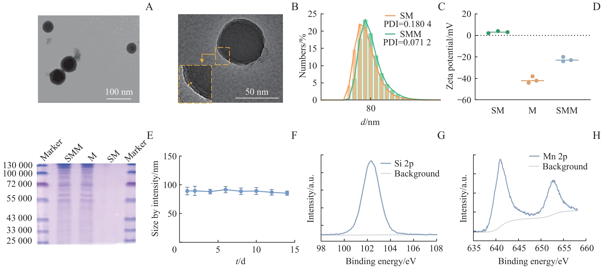
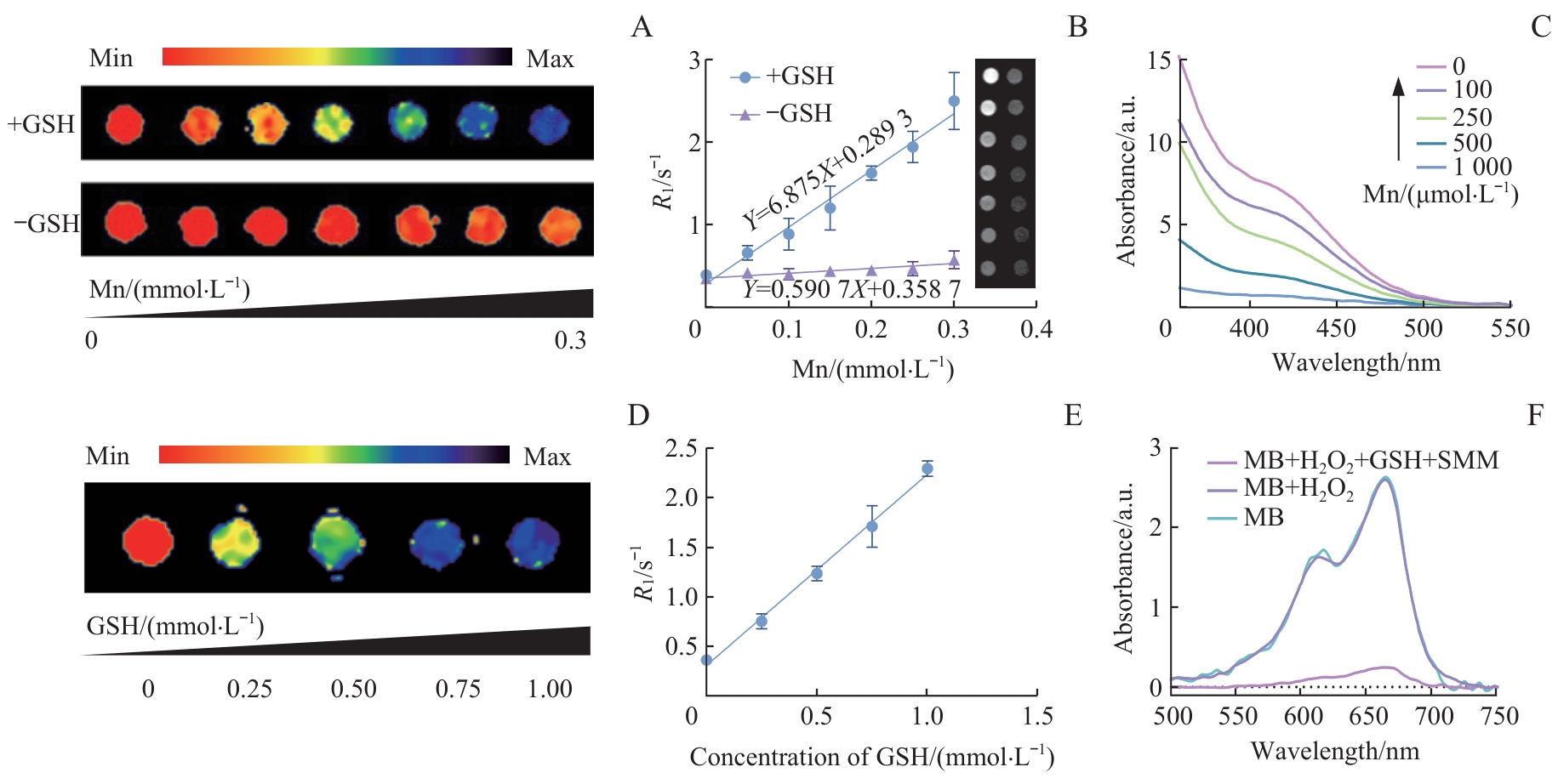
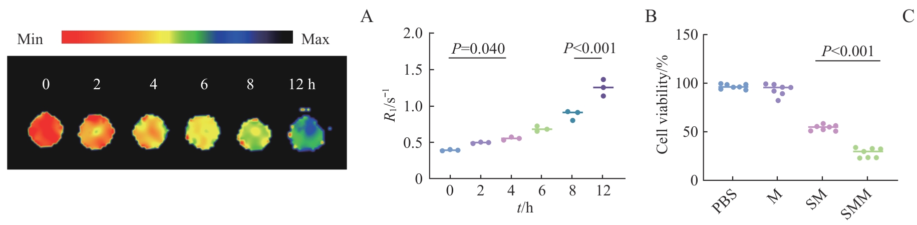
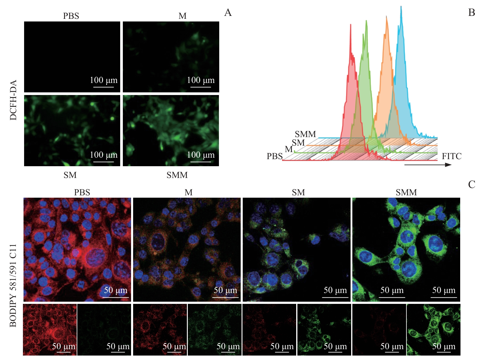
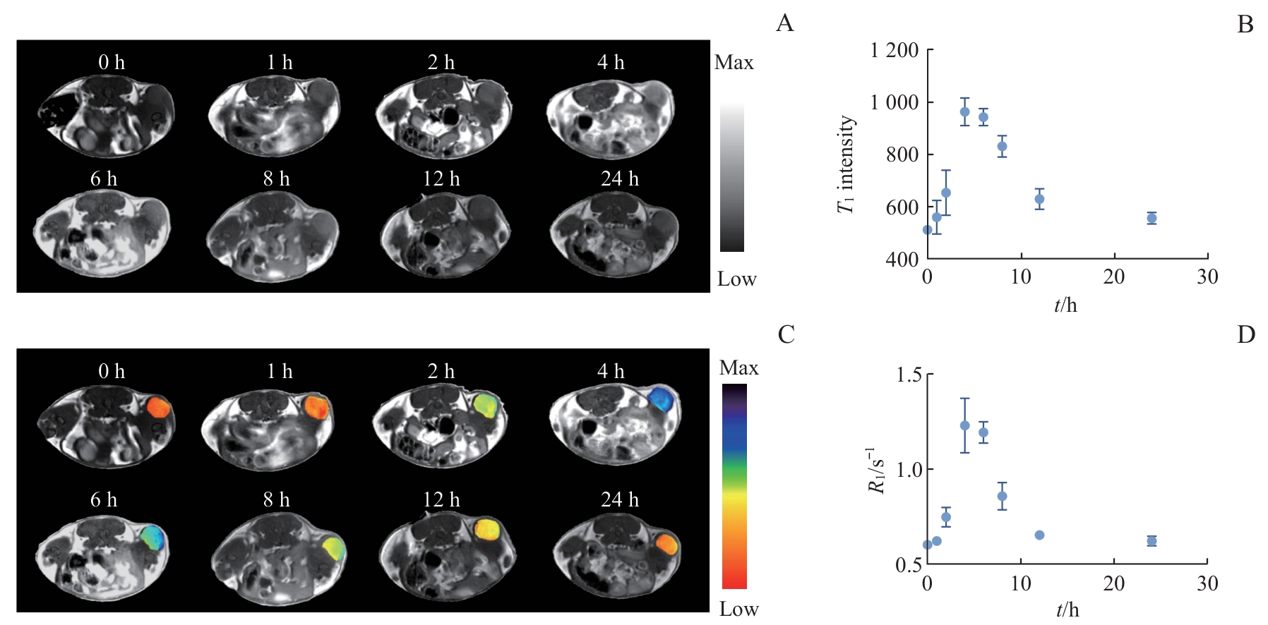
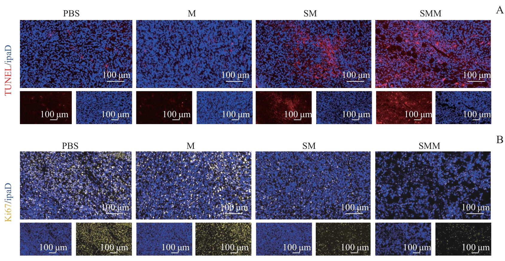
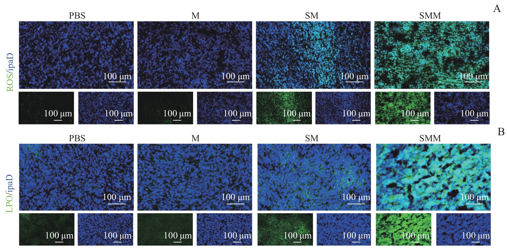
目的·构建一种细胞膜仿生、谷胱甘肽(glutathione,GSH)响应性的锰基纳米材料诊疗探针,并探究其生物膜靶向能力、通过GSH耗竭诱导三阴性乳腺癌(triple-negative breast cancer,TNBC)铁死亡效能,以及磁共振(magnetic resonance,MR)激活成像的实时监测效应。方法·采用溶胶-凝胶法制备二氧化硅(SiO2)纳米核心,借助氧化还原共沉淀法在其表面沉积二氧化锰(MnO2),最后利用薄膜挤压法包覆小鼠4T1乳腺癌细胞膜,从而合成仿生纳米探针SiO2@MnO2@Membrane(SMM)。采用透射电子显微镜(transmission electron microscope,TEM)与粒径仪对其形貌、粒径及电位进行表征。采用考马斯亮蓝染色法验证材料表面的细胞膜蛋白负载情况;采用X射线光电子能谱(X- ray photoelectron spectroscopy,XPS)表征材料中的特征元素。通过MR成像评估SMM的GSH响应性T1信号激活效能。利用亚甲基蓝及5,5′-二硫双(2-硝基苯甲酸)[5,5′-dithiobis(2-nitrobenzoic acid),DTNB]分别检测无细胞体系中活性氧(reactive oxygen species,ROS)的生成及GSH的消耗情况。采用噻唑蓝法评估纳米探针对4T1乳腺癌细胞的杀伤效能。借助2′,7′-二氯二氢荧光素(2′,7′-dichlorodihydrofluorescein diacetate,DCFH-DA)和BODIPY 581/591 C11探针检测细胞内ROS生成和脂质过氧化物(lipid peroxides,LPO)的累积水平。构建小鼠4T1 TNBC皮下移植瘤模型,通过MR动态监测探针在肿瘤部位的T1激活效应,并结合肿瘤组织中LPO、ROS等铁死亡标志物,以及细胞增殖核抗原(Ki-67)、脱氧核糖核苷酸末端转移酶介导的缺口末端标记法(terminal-deoxynucleotidyl transferase mediated nick end labeling,TUNEL)的检测结果,评估其基于铁死亡的抗肿瘤效能。结果·TEM显示,制备的SMM纳米探针呈规则球形结构。粒径仪检测表明,包覆细胞膜后,材料的Zeta电位从(+3.2±1.0)mV转变为(-22.0±2.0)mV,粒径从68 nm增加至78 nm,多分散性指数(polydispersity index,PDI)维持在较低水平,呈现均一稳定状态。考马斯亮蓝染色验证了生物膜的有效包覆。XPS检测到材料中Mn和Si的特征元素信号。上述结果共同证实SMM纳米探针成功合成。MR结果显示,SMM在GSH存在时可显著激活T1信号,纵向摩尔弛豫率(r1)由0.591 (mmol/L)-1·s-1显著提升至6.875 (mmol/L)-1·s-1;细胞水平的T1信号呈时间依赖性激活。DCFH-DA与BODIPY 581/591 C11染色结果显示,SMM组细胞内大量累积ROS与LPO,且细胞存活率较对照组显著降低(28.35%±4.91%)。体内实验结果显示,尾静脉注射SMM后,肿瘤区域T1信号在4 h达到峰值(1.23±0.14)s-1,表明探针可靶向富集于TNBC肿瘤组织并激活MR T1信号。肿瘤组织切片检测显示,SMM组ROS及LPO水平显著升高,同时伴随Ki-67表达下调、TUNEL阳性信号上调,进一步证实其可通过诱导铁死亡发挥抗肿瘤作用。结论·成功构建了基于GSH耗竭的4T1细胞膜仿生纳米探针SMM。该探针能够在TNBC肿瘤部位特异性激活T1信号,同时通过靶向富集协同诱导肿瘤细胞铁死亡,最终显著提升抗肿瘤疗效。

关键词: 铁死亡, 细胞膜仿生, 三阴性乳腺癌, 磁共振成像, 锰基纳米粒, 谷胱甘肽耗竭, 活性氧

Abstract:

Objective ·To develop a biomimetic membrane, glutathione (GSH)-responsive manganese-based theranostic nanoprobe, and investigate its biomembrane-targeting capability, efficacy in GSH-depletion-mediated ferroptosis induction in triple-negative breast cancer (TNBC), and real-time monitoring of MR signal activation. Methods ·A silica (SiO2) nanoparticle core was synthesized via the sol-gel method, followed by deposition of manganese dioxide (MnO2) onto its surface through redox co-precipitation, and then coating with 4T1 breast cancer cell membranes using an extrusion technique to yield the biomimetic nanoprobe SiO2@MnO2@Membrane (SMM). The morphology, particle size, and Zeta potential were characterized by transmission electron microscopy (TEM) and dynamic light scattering (DLS). Membrane protein retention on the nanoparticle surface was confirmed by Coomassie brilliant blue staining, and characteristic elements was analyzed by X-ray photoelectron spectroscopy (XPS). The GSH-responsive T1 MR signal activation was evaluated in vitro. Reactive oxygen species (ROS) generation and GSH depletion in a cell-free system were assessed using methylene blue and 5, 5′-dithiobis (2-nitrobenzoic acid) (DTNB) assays, respectively. The cytotoxic effect of the nanoprobe on 4T1 cells was determined by the MTT assay. Intracellular ROS generation and lipid peroxide (LPO) accumulation were detected using 2′,7′-dichlorodihydrofluoresce in diacetate (DCFH-DA) and BODIPY 581/591 C11 probes. A murine subcutaneous TNBC model was established to dynamically monitor T1-weighted MR signal activation of the nanoprobe at tumor sites. Combined with the detection of ferroptosis-related markers, including LPO and ROS, as well as Ki-67 and terminal-deoxynucleotidyl transferase-mediated nick end labeling (TUNEL) staining in tumor tissues, the ferroptosis-mediated antitumor efficacy of SMM was evaluated. Results ·TEM revealed that the prepared SMM nanoprobe exhibited a regular spherical structure. Particle size analysis showed that after cell membrane coating, the Zeta potential shifted from (+3.2±1.0) mV to (-22.0±2.0) mV, and the hydrodynamic diameter increased from 68 nm to 78 nm, while the polydisper sity index (PDI) remained at a low level, indicating a uniform and stable dispersion. Coomassie brilliant blue staining confirmed effective biomimetic membrane coating. XPS detected characteristic signals of Mn and Si elements, and these results collectively confirmed the successful synthesis of the SMM nanoprobe. MR results showed that SMM significantly activated the T1 signal in the presence of GSH, with relaxivity (r1). increasing significantly from 0.591 to 6.875 (mmol/L)-1·s-1. At the cellular level, T1 signal activation exhibited a time-dependent enhancement. Compared to the control groups, DCFH-DA and BODIPY 581/591 C11 staining indicated substantial intracellular accumulation of ROS and LPO in the SMM group, accompanied by a significant reduction in cell viability (28.35%±4.91% survival). In vivo experiments showed that after intravenous injection of SMM,the T1 signal intensity in tumor regions reached a peak value of (1.23±0.14) s-1 at 4 h, indicating that the nanoprobe could specifically accumulate in TNBC tumor tissues and activate MR T1 signals. Tumor tissue analysis revealed significantly elevated ROS and LPO levels in the SMM group, accompanied by decreased Ki-67 expression and increased TUNEL-positive signals, further confirming its antitumor effect through ferroptosis induction. Conclusion ·A GSH-depleting 4T1 cell membrane-biomimetic nanoprobe (SMM) was successfully developed, which can specifically activate T1 MR signals at TNBC tumor sites and synergistically induce ferroptosis through targeted accumulation, significantly enhancing antitumor efficacy.

Key words: ferroptosis, biomimetic membrane, triple-negative breast cancer, magnetic resonance imaging, manganese-based nanoparticle, glutathione depletion, reactive oxygen species

中图分类号: