上海交通大学学报(医学版) ›› 2022, Vol. 42 ›› Issue (9): 1311-1314.doi: 10.3969/j.issn.1674-8115.2022.09.018

• 论著 · 临床研究 • 上一篇    

1型糖尿病儿童眼底黄斑区早期微血管变化的观察

李雯1(), 李苑1, 叶海昀1, 张笑笑1, 乔彤1(), 李嫔2   

  1. 1.上海市儿童医院,上海交通大学医学院附属儿童医院眼科,上海 200062
    2.上海市儿童医院,上海交通大学医学院附属儿童医院内分泌科,上海 200062
  • 收稿日期:2022-05-27 接受日期:2022-07-23 出版日期:2022-09-28 发布日期:2022-09-28
  • 通讯作者: 乔彤 E-mail:liwen458@sina.com;qiaot@shchildren.com.cn
  • 作者简介:李 雯(1976—),女,副主任医师,硕士;电子信箱:liwen458@sina.com
  • 基金资助:
    上海市“科技创新行动计划”医学创新研究专项项目(20Z11900400)

Observation on early microvascular changes in macular area of the fundus in children with type 1 diabetes mellitus

LI Wen1(), LI Yuan1, YE Haiyun1, ZHANG Xiaoxiao1, QIAO Tong1(), LI Pin2   

  1. 1.Department of Ophthalmology, Shanghai Children's Hospital, Shanghai Jiao Tong University School of Medicine, Shanghai 200062, China
    2.Department of Endocrinology, Shanghai Children's Hospital, Shanghai Jiao Tong University School of Medicine, Shanghai 200062, China
  • Received:2022-05-27 Accepted:2022-07-23 Online:2022-09-28 Published:2022-09-28
  • Contact: QIAO Tong E-mail:liwen458@sina.com;qiaot@shchildren.com.cn
  • Supported by:
    “Science and Technology Innovation Action Plan” Medical Innovation Research Special Project of Shanghai(20Z11900400)

摘要:

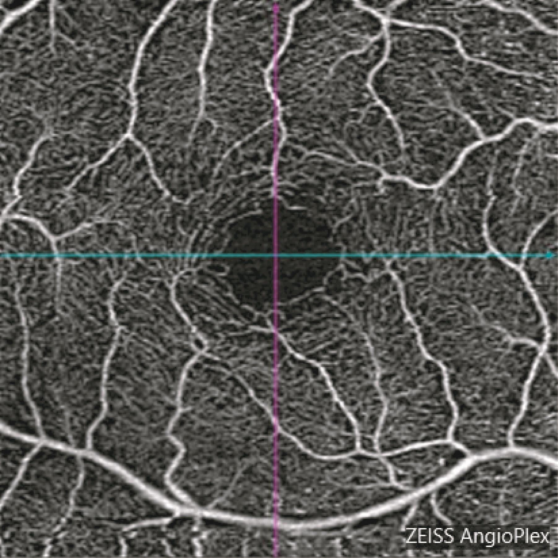
目的·利用光学相干断层扫描血管成像技术(optical coherence tomography angiography,OCTA)观察尚未出现视网膜病变的1型糖尿病儿童眼底黄斑区血管形态及血流变化,以及探索OCTA在该类儿童中的应用效果。方法·选择2019年6月—2020年2月于上海交通大学医学院附属儿童医院内分泌科住院的无视网膜病变的1型糖尿病儿童27例(54只眼,为观察组),及与其年龄相匹配的健康儿童25例(50只眼,为对照组)。采用OCTA对2组儿童的眼底黄斑区分别行3 mm×3 mm、6 mm×6 mm范围扫描,观察其黄斑拱环形态,并对黄斑中心凹无血管区(foveal avascular zone,FAZ)面积、中心凹周围不同分区的血管密度及灌注密度进行定量分析。结果·观察组儿童的眼底黄斑拱环形态较规则,其黄斑FAZ面积较对照组扩大(P=0.000),其黄斑中心凹周围直径1 mm范围内浅层视网膜血管密度及灌注密度均较对照组降低(P=0.009,P=0.012)。而该组儿童的眼底黄斑中心凹周围直径1 mm外、直径3 mm内区域的浅层视网膜血管密度为(18.29±0.96)mm-1、灌注密度为0.43±0.03,黄斑中心凹周围直径3 mm外、直径6 mm内区域的浅层视网膜血管密度为(18.58±0.69)mm-1、灌注密度为0.46±0.02,与对照组间差异均无统计学意义。结论·1型糖尿病儿童出现视网膜病变之前,已存在黄斑拱环的扩大、黄斑中心凹周围1 mm直径范围内浅层视网膜血管密度及灌注密度的下降。OCTA能对该类儿童眼底黄斑区血管形态及血流变化进行有效的早期监测。

关键词: 光学相干断层扫描血管成像技术, 糖尿病视网膜病变, 1型糖尿病, 儿童

Abstract:

Objective ·To observe the changes of vascular morphology and blood flow in the macular area of the fundus of type 1 diabetic children without retinopathy by optical coherence tomography angiography (OCTA), and explore the application of OCTA in these children. Methods ·From June 2019 to February 2020, 27 type 1 diabetic children without retinopathy (54 eyes, observation group) who were hospitalized in the Department of Endocrinology of Shanghai Children′s Hospital, Shanghai Jiao Tong University School of Medicine, and 25 age-matched healthy children (50 eyes, control group) were selected. The macular area of the fundus of the two groups of children was scanned with OCTA in the range of 3 mm×3 mm and 6 mm×6 mm, respectively, and the morphology of the macular arch ring was observed. The area of foveal avascular zone (FAZ), and the vascular density and perfusion density of different zones around the foveal were quantitatively analyzed Results ·The morphology of the macular arch ring in the observation group was regular, and the FAZ area of macular area was larger than that in the control group (P=0.000). The superficial retinal vascular density and perfusion density within the diameter of 1 mm around the macular fovea were lower than those in the control group (P=0.009, P=0.012). In the observation group, the superficial retinal vascular density and perfusion density in the area outside 1 mm diameter and within 3 mm diameter around the macular fovea were (18.29±0.96) mm-1 and 0.43±0.03, while the superficial retinal vascular density and perfusion density in the area outside 3 mm diameter and within 6 mm diameter around the macular fovea were (18.58±0.69) mm-1 and 0.46±0.02, which had no significant difference with the control group. Conclusion ·In the children with type 1 diabetes mellitus, the enlargement of macular arch and the decrease of superficial retinal vascular density and perfusion density within 1 mm diameter around macular fovea may occur before retinopathy. OCTA can be effectively applied to the early monitoring of the changes of vascular morphology and blood flow in the macular area of the fundus of diabetic children.

Key words: optical coherence tomography angiography (OCTA), diabetic retinopathy (DR), type 1 diabetes mellitus, child

中图分类号: