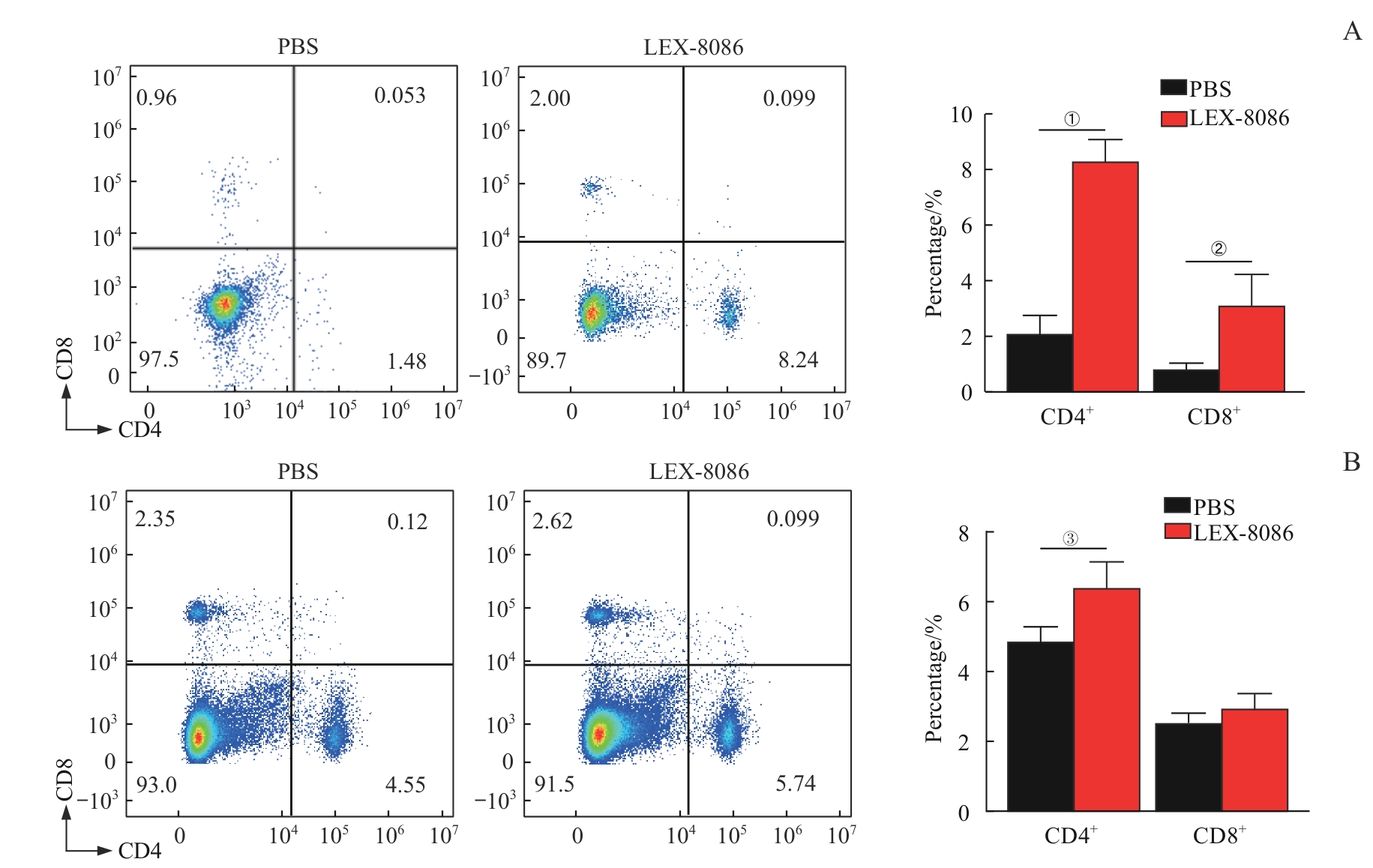
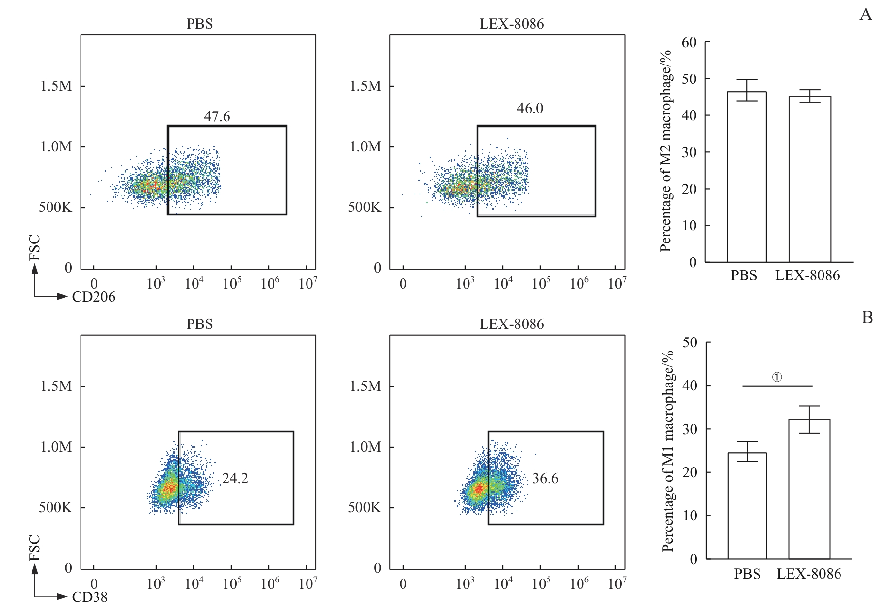
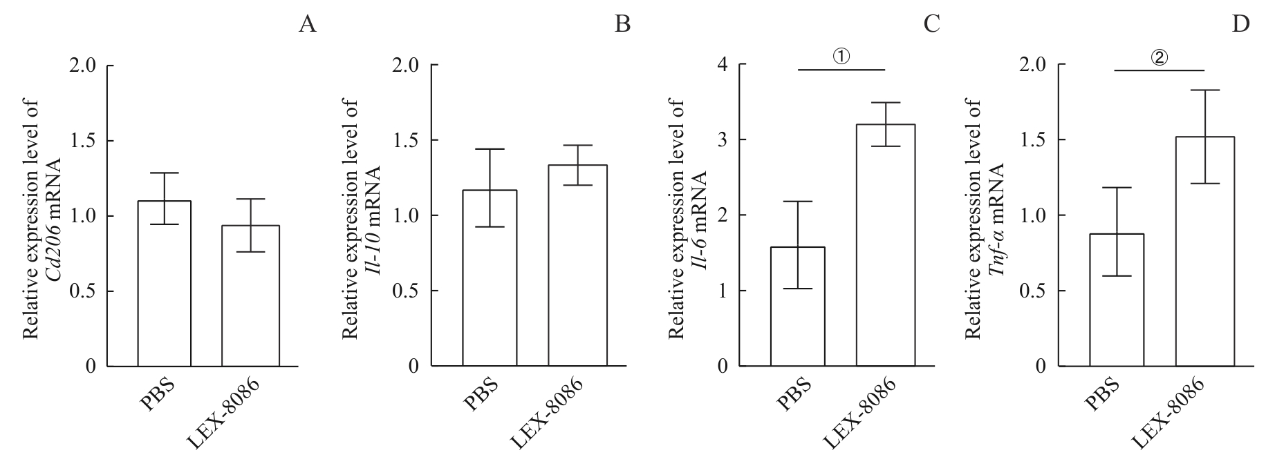
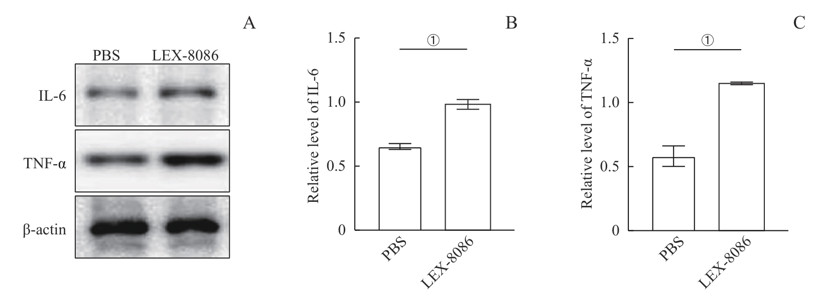
| 1 |
WHITESIDE T L. Tumor-derived exosomes and their role in cancer progression[J]. Adv Clin Chem, 2016, 74: 103-141.
|
| 2 |
SUN Z Y, XIE C Q, LIU H, et al. CD19 CAR-T cell therapy induced immunotherapy associated interstitial pneumonitis: a case report[J]. Front Immunol, 2022, 13: 778192.
|
| 3 |
RAJE N, BERDEJA J, LIN Y, et al. Anti-BCMA CAR T-cell therapy bb2121 in relapsed or refractory multiple myeloma[J]. N Engl J Med, 2019, 380(18): 1726-1737.
|
| 4 |
OTT P A, HU Z T, KESKIN D B, et al. An immunogenic personal neoantigen vaccine for patients with melanoma[J]. Nature, 2017, 547(7662): 217-221.
|
| 5 |
SHEIH A, VOILLET V, HANAFI L A, et al. Clonal kinetics and single-cell transcriptional profiling of CAR-T cells in patients undergoing CD19 CAR-T immunotherapy[J]. Nat Commun, 2020, 11(1): 219.
|
| 6 |
CHEN G, HUANG A C, ZHANG W, et al. Exosomal PD-L1 contributes to immunosuppression and is associated with anti-PD-1 response[J]. Nature, 2018, 560(7718): 382-386.
|
| 7 |
AHMAD A. Epigenetic regulation of immunosuppressive tumor-associated macrophages through dysregulated microRNAs[J]. Semin Cell Dev Biol, 2022, 124: 26-33.
|
| 8 |
NASERI M, BOZORGMEHR M, ZÖLLER M, et al. Tumor-derived exosomes: the next generation of promising cell-free vaccines in cancer immunotherapy[J]. Oncoimmunology, 2020, 9(1): 1779991.
|
| 9 |
KRACKHARDT A M, HARIG S, WITZENS M, et al. T-cell responses against chronic lymphocytic leukemia cells: implications for immunotherapy[J]. Blood, 2002, 100(1): 167-173.
|
| 10 |
TOWNSEND S E, ALLISON J P. Tumor rejection after direct costimulation of CD8+ T cells by B7-transfected melanoma cells[J]. Science, 1993, 259(5093): 368-370.
|
| 11 |
CHENG Q Z, KANG Y, YAO B, et al. Genetically engineered-cell-membrane nanovesicles for cancer immunotherapy[J]. Adv Sci (Weinh), 2023, 10(26): e2302131.
|
| 12 |
HU W W, HUANG F, NING L X, et al. Enhanced immunogenicity of leukemia-derived exosomes via transfection with lentiviral vectors encoding costimulatory molecules[J]. Cell Oncol (Dordr), 2020, 43(5): 889-900.
|
| 13 |
LI J Q, HUANG F, JIANG Y, et al. A novel costimulatory molecule gene-modified leukemia cell-derived exosome-targeted CD4+ T cell vaccine efficiently enhances anti-leukemia immunity[J]. Front Immunol, 2022, 13: 1043484.
|
| 14 |
JOHNSON B D, YAN X C, SCHAUER D W, et al. Dual expression of CD80 and CD86 produces a tumor vaccine superior to single expression of either molecule[J]. Cell Immunol, 2003, 222(1): 15-26.
|
| 15 |
VASILEVKO V, GHOCHIKYAN A, HOLTERMAN M J, et al. CD80 (B7-1) and CD86 (B7-2) are functionally equivalent in the initiation and maintenance of CD4+ T-cell proliferation after activation with suboptimal doses of PHA[J]. DNA Cell Biol, 2002, 21(3): 137-149.
|
| 16 |
SIVORI S, PENDE D, QUATRINI L, et al. NK cells and ILCs in tumor immunotherapy[J]. Mol Aspects Med, 2021, 80: 100870.
|
| 17 |
TRACY S I, VENKATESH H, HEKIM C, et al. Combining nilotinib and PD-L1 blockade reverses CD4+ T-cell dysfunction and prevents relapse in acute B-cell leukemia[J]. Blood, 2022, 140(4): 335-348.
|
| 18 |
DISTLER E, ALBRECHT J, BRUNK A, et al. Patient-individualized CD8⁺ cytolytic T-cell therapy effectively combats minimal residual leukemia in immunodeficient mice[J]. Int J Cancer, 2016, 138(5): 1256-1268.
|
 ), 王明慧, 赵洁, 万江波, 黄方(