上海交通大学学报(医学版) ›› 2025, Vol. 45 ›› Issue (4): 500-507.doi: 10.3969/j.issn.1674-8115.2025.04.013
张欣欣1, 颜崇淮1,2( )
)
收稿日期:2024-11-10
接受日期:2024-12-19
出版日期:2025-04-28
发布日期:2025-04-21
通讯作者:
颜崇淮,主任医师,博士;电子信箱:yanchonghuai@xinhuamed.com.cn。作者简介:张欣欣(1999—),女,硕士生;电子信箱:beverlyz0201@163.com。
基金资助:
ZHANG Xinxin1, YAN Chonghuai1,2()
Received:2024-11-10
Accepted:2024-12-19
Online:2025-04-28
Published:2025-04-21
Contact:
YAN Chonghuai, E-mail: yanchonghuai@xinhuamed.com.cn.Supported by:摘要:
铅是一种普遍存在于环境中的有毒重金属,也是人类历史上使用最早、应用最为广泛的重金属元素之一。由于铅在环境中不可降解,并且在人体内具有较长的生物累积效应(可长达30~50年),即使极低浓度的铅也能对人体造成健康损害,因而被世界卫生组织(World Health Organization,WHO)列为十大公共卫生关注化学品之一。铅进入人体后,通常会分布在脑、肝脏、肾脏、牙齿和骨骼等组织中,进而对全身各个系统、多种脏器和组织产生广泛的毒性作用。表观遗传学是研究基因表达在不改变核苷酸序列的情况下发生可遗传变化的学科,它揭示了基因表达修饰如何对细胞进行调控,导致具有相同DNA序列的细胞表现出不同形态与功能。尽管铅的毒性机制尚未完全明确,但近年来的研究表明,表观遗传学调控可能是铅毒性作用的重要机制之一。环境铅暴露可通过引发个体细胞的DNA甲基化、组蛋白修饰和微RNA(microRNA,miRNA)等表观遗传学改变,进而而诱发多种毒性反应。该文就铅毒性相关的表观遗传学机制研究现状,着重从DNA甲基化、组蛋白修饰和miRNA 3个方面进行综述,旨在从表观遗传学角度审视铅毒性,并为进一步探究铅的毒性机制提供理论基础。
中图分类号:
张欣欣, 颜崇淮. 铅毒性的表观遗传学机制研究进展[J]. 上海交通大学学报(医学版), 2025, 45(4): 500-507.
ZHANG Xinxin, YAN Chonghuai. Advances in epigenetic mechanisms of lead toxicity[J]. Journal of Shanghai Jiao Tong University (Medical Science), 2025, 45(4): 500-507.
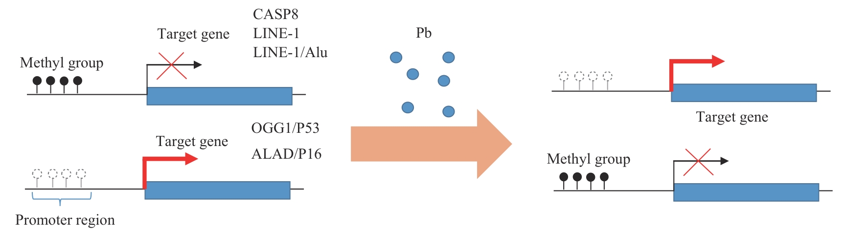
图1 铅对DNA甲基化的调控以及对下游基因的两面性
Fig 1 Dual effects of lead on DNA methylation and downstream genes
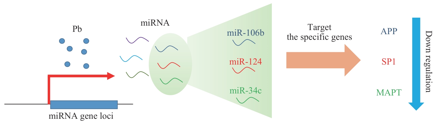
图2 铅暴露诱导AD相关miRNA变化并抑制特定基因表达
Fig 2 Lead Exposure alters AD-related miRNAs and suppresses gene expression
| Gene | Expression change | Sample type | Effect | Reference |
|---|---|---|---|---|
| miR-155/miR-221 | Upregulated | Peripheral blood samples from adults | Cell apoptosis, cell proliferation, and cytokine production | PMID: 32902755 |
| miR-146a | Upregulated | Peripheral blood samples from adults | Inhibition of inflammatory factor release and cell apoptosis | PMID: 36274319 |
| miR-106b-56p | Upregulated | HT-22 and PC12 cell lines | Decreased XIAP levels and cell viability | PMID: 32344020 |
| miR-378a-3p | Upregulated | HT-22 cell line | Reduced GSH and increased lipid ROS levels | PMID: 35588615 |
| miR-143-5p | Downregulated | Fibroblast samples | Regulation of dysfunction of interstitial fibroblasts | PMID: 35485286 |
| miR-106b/miR-124/miR-34c | Upregulated | Cerebral cortex tissue samples | Induced neurotoxicity and learning/memory impairment | PMID: 29614648/27293183 |
| miR-34b | Upregulated | Hippocampal tissue samples | Induced developmental neuropsychiatric dysfunction | PMID: 35367965 |
表1 铅暴露对miRNA的调控及其相关效应
Tab 1 Regulation of miRNA by Pb exposure and its related effects
| Gene | Expression change | Sample type | Effect | Reference |
|---|---|---|---|---|
| miR-155/miR-221 | Upregulated | Peripheral blood samples from adults | Cell apoptosis, cell proliferation, and cytokine production | PMID: 32902755 |
| miR-146a | Upregulated | Peripheral blood samples from adults | Inhibition of inflammatory factor release and cell apoptosis | PMID: 36274319 |
| miR-106b-56p | Upregulated | HT-22 and PC12 cell lines | Decreased XIAP levels and cell viability | PMID: 32344020 |
| miR-378a-3p | Upregulated | HT-22 cell line | Reduced GSH and increased lipid ROS levels | PMID: 35588615 |
| miR-143-5p | Downregulated | Fibroblast samples | Regulation of dysfunction of interstitial fibroblasts | PMID: 35485286 |
| miR-106b/miR-124/miR-34c | Upregulated | Cerebral cortex tissue samples | Induced neurotoxicity and learning/memory impairment | PMID: 29614648/27293183 |
| miR-34b | Upregulated | Hippocampal tissue samples | Induced developmental neuropsychiatric dysfunction | PMID: 35367965 |
| 1 | MEI Z Q, LIU G F, ZHAO B, et al. Emerging roles of epigenetics in lead-induced neurotoxicity[J]. Environ Int, 2023, 181: 108253. |
| 2 | APPLETON A A, JACKSON B P, KARAGAS M, et al. Prenatal exposure to neurotoxic metals is associated with increased placental glucocorticoid receptor DNA methylation[J]. Epigenetics, 2017, 12(8): 607-615. |
| 3 | RECILLAS-TARGA F. Cancer epigenetics: an overview[J]. Arch Med Res, 2022, 53(8): 732-740. |
| 4 | OKAMOTO Y, IWAI-SHIMADA M, NAKAI K, et al. Global DNA methylation in cord blood as a biomarker for prenatal lead and antimony exposures[J]. Toxics, 2022, 10(4): 157. |
| 5 | ZENG Z J, HUO X, ZHANG Y, et al. Differential DNA methylation in newborns with maternal exposure to heavy metals from an e-waste recycling area[J]. Environ Res, 2019, 171: 536-545. |
| 6 | BOZACK A K, RIFAS-SHIMAN S L, COULL B A, et al. Prenatal metal exposure, cord blood DNA methylation and persistence in childhood: an epigenome-wide association study of 12 metals[J]. Clin Epigenetics, 2021, 13(1): 208. |
| 7 | TUNG P W, KENNEDY E M, BURT A, et al. Prenatal lead (Pb) exposure is associated with differential placental DNA methylation and hydroxymethylation in a human population[J]. Epigenetics, 2022, 17(13): 2404-2420. |
| 8 | BRAUN J M. Early-life exposure to EDCs: role in childhood obesity and neurodevelopment[J]. Nat Rev Endocrinol, 2017, 13(3): 161-173. |
| 9 | RYGIEL C A, DOLINOY D C, PERNG W, et al. Trimester-specific associations of prenatal lead exposure with infant cord blood DNA methylation at birth[J]. Epigenet Insights, 2020, 13: 2516865720938669. |
| 10 | RYGIEL C A, DOLINOY D C, BAKULSKI K M, et al. DNA methylation at birth potentially mediates the association between prenatal lead (Pb) exposure and infant neurodevelopmental outcomes[J]. Environ Epigenet, 2021, 7(1): dvab005. |
| 11 | WANG K, LIU S Y, SVOBODA L K, et al. Tissue- and sex-specific DNA methylation changes in mice perinatally exposed to lead (Pb)[J]. Front Genet, 2020, 11: 840. |
| 12 | SVOBODA L K, NEIER K R, WANG K, et al. Tissue and sex-specific programming of DNA methylation by perinatal lead exposure: implications for environmental epigenetics studies[J]. Epigenetics, 2021, 16(10): 1102-1122. |
| 13 | SVOBODA L K, WANG K, JONES T R, et al. Sex-specific alterations in cardiac DNA methylation in adult mice by perinatal lead exposure[J]. Int J Environ Res Public Health, 2021, 18(2): 577. |
| 14 | SVOBODA L K, WANG K, GOODRICH J M, et al. Perinatal lead exposure promotes sex-specific epigenetic programming of disease-relevant pathways in mouse heart[J]. Toxics, 2023, 11(1): 85. |
| 15 | MORGAN R K, WANG K, SVOBODA L K, et al. Effects of developmental lead and phthalate exposures on DNA methylation in adult mouse blood, brain, and liver identifies tissue- and sex-specific changes with implications for genomic imprinting[J]. bioRxiv, 2023: 2023.09.29.560131. |
| 16 | MCCABE C, ANDERSON O S, MONTROSE L, et al. Sexually dimorphic effects of early-life exposures to endocrine disruptors: sex-specific epigenetic reprogramming as a potential mechanism[J]. Curr Environ Health Rep, 2017, 4(4): 426-438. |
| 17 | WARKOCKI Z. An update on post-transcriptional regulation of retrotransposons[J]. FEBS Lett, 2023, 597(3): 380-406. |
| 18 | WANG K, MENG Y, WANG T W, et al. Global and gene-specific promoter methylation, and micronuclei induction in lead-exposed workers: a cross-sectional study[J]. Environ Mol Mutagen, 2021, 62(7): 428-434. |
| 19 | WANG T W, MENG Y, TU Y T, et al. Associations between DNA methylation and genotoxicity among lead-exposed workers in China[J]. Environ Pollut, 2023, 316(Pt 1): 120528. |
| 20 | EL-SHETRY E S, MOHAMED A A, KHATER S I, et al. Synergistically enhanced apoptotic and oxidative DNA damaging pathways in the rat brain with lead and/or aluminum metals toxicity: expression pattern of genes OGG1 and P53[J]. J Trace Elem Med Biol, 2021, 68: 126860. |
| 21 | YOHANNES Y B, NAKAYAMA S M, YABE J, et al. Blood lead levels and aberrant DNA methylation of the ALAD and p16 gene promoters in children exposed to environmental-lead[J]. Environ Res, 2020, 188: 109759. |
| 22 | CARDELLI M. The epigenetic alterations of endogenous retroelements in aging[J]. Mech Ageing Dev, 2018, 174: 30-46. |
| 23 | GOKHMAN D, MALUL A, CARMEL L. Inferring past environments from ancient epigenomes[J]. Mol Biol Evol, 2017, 34(10): 2429-2438. |
| 24 | COLICINO E, JUST A, KIOUMOURTZOGLOU M A, et al. Blood DNA methylation biomarkers of cumulative lead exposure in adults[J]. J Expo Sci Environ Epidemiol, 2021, 31(1): 108-116. |
| 25 | PAUL K C, HORVATH S, DEL ROSARIO I, et al. DNA methylation biomarker for cumulative lead exposure is associated with Parkinson's disease[J]. Clin Epigenetics, 2021, 13(1): 59. |
| 26 | LIEBERMAN-CRIBBIN W, DOMINGO-RELLOSO A, NAVAS-ACIEN A, et al. Epigenetic biomarkers of lead exposure and cardiovascular disease: prospective evidence in the strong heart study[J]. J Am Heart Assoc, 2022, 11(23): e026934. |
| 27 | HOCHER A, WARNECKE T. Nucleosomes at the dawn of eukaryotes[J]. Genome Biol Evol, 2024, 16(3): evae029. |
| 28 | SÁNCHEZ O F, LIN L F, XIE J K, et al. Lead exposure induces dysregulation of constitutive heterochromatin hallmarks in live cells[J]. Curr Res Toxicol, 2022, 3: 100061. |
| 29 | GU X Z, XU Y, XUE W Z, et al. Interplay of miR-137 and EZH2 contributes to the genome-wide redistribution of H3K27me3 underlying the Pb-induced memory impairment[J]. Cell Death Dis, 2019, 10(9): 671. |
| 30 | XIAO J, WANG T, XU Y, et al. Long-term probiotic intervention mitigates memory dysfunction through a novel H3K27me3-based mechanism in lead-exposed rats[J]. Transl Psychiatry, 2020, 10(1): 25. |
| 31 | EID A, BIHAQI S W, RENEHAN W E, et al. Developmental lead exposure and lifespan alterations in epigenetic regulators and their correspondence to biomarkers of Alzheimer's disease[J]. Alzheimers Dement (Amst), 2016, 2: 123-131. |
| 32 | VARMA G, SOBOLEWSKI M, CORY-SLECHTA D A, et al. Sex- and brain region- specific effects of prenatal stress and lead exposure on permissive and repressive post-translational histone modifications from embryonic development through adulthood[J]. Neurotoxicology, 2017, 62: 207-217. |
| 33 | SNIGDHA S, ALEPH PRIETO G, PETROSYAN A, et al. H3K9me3 inhibition improves memory, promotes spine formation, and increases BDNF levels in the aged hippocampus[J]. J Neurosci, 2016, 36(12): 3611-3622. |
| 34 | LIN L F, XIE J K, SÁNCHEZ O F, et al. Low dose lead exposure induces alterations on heterochromatin hallmarks persisting through SH-SY5Y cell differentiation[J]. Chemosphere, 2021, 264(Pt 1): 128486. |
| 35 | KUMAR K, ANJALI S, SHARMA S. Effect of lead exposure on histone modifications: a review[J]. J Biochem Mol Toxicol, 2024, 38(1): e23547. |
| 36 | LUO M, XU Y, CAI R, et al. Epigenetic histone modification regulates developmental lead exposure induced hyperactivity in rats[J]. Toxicol Lett, 2014, 225(1): 78-85. |
| 37 | XU L H, MU F F, ZHAO J H, et al. Lead induces apoptosis and histone hyperacetylation in rat cardiovascular tissues[J]. PLoS One, 2015, 10(6): e0129091. |
| 38 | KIRAN G S, KUMAR P K, MITRA P, et al. Unravelling blood-based epigenetic mechanisms: the impact of hsa-miR-146a and histone H3 acetylation in lead-induced inflammation among occupational workers[J]. Int Arch Occup Environ Health, 2023, 96(9): 1257-1266. |
| 39 | WANG Y W, HU Y Z, WU Z T, et al. Latent role of in vitro Pb exposure in blocking Aβ clearance and triggering epigenetic modifications[J]. Environ Toxicol Pharmacol, 2019, 66: 14-23. |
| 40 | WU Y L, XU Y, HUANG X Y, et al. Regulatory roles of histone deacetylases 1 and 2 in Pb-induced neurotoxicity[J]. Toxicol Sci, 2018, 162(2): 688-701. |
| 41 | CHESHMAZAR N, HAMZEH-MIVEHROUD M, NOZAD CHAROUDEH H, et al. Current trends in development of HDAC-based chemotherapeutics[J]. Life Sci, 2022, 308: 120946. |
| 42 | ZHOU R Q, ZHAO J, LI D Y, et al. Combined exposure of lead and cadmium leads to the aggravated neurotoxicity through regulating the expression of histone deacetylase 2[J]. Chemosphere, 2020, 252: 126589. |
| 43 | GU X Z, HUANG X Y, LI D Y, et al. Nuclear accumulation of histone deacetylase 4 (HDAC4) by PP1-mediated dephosphorylation exerts neurotoxicity in Pb-exposed neural cells[J]. Neurotoxicology, 2020, 81: 395-405. |
| 44 | GU X Z, SHEN N, HUANG C Q, et al. Pb inhibited C2C12 myoblast differentiation by regulating HDAC2[J]. Toxicology, 2023, 499: 153639. |
| 45 | XU M, YU Z M, HU F F, et al. Identification of differential plasma miRNA profiles in Chinese workers with occupational lead exposure[J]. Biosci Rep, 2017, 37(5): BSR20171111. |
| 46 | OCHOA-MARTÍNEZ Á C, VARELA-SILVA J A, ORTA-GARCÍA S T, et al. Lead (Pb) exposure is associated with changes in the expression levels of circulating miRNAS (miR-155, miR-126) in Mexican women[J]. Environ Toxicol Pharmacol, 2021, 83: 103598. |
| 47 | WEN Q F, VERHEIJEN M, WITTENS M M J, et al. Lead-exposure associated miRNAs in humans and Alzheimer's disease as potential biomarkers of the disease and disease processes[J]. Sci Rep, 2022, 12(1): 15966. |
| 48 | MITRA P, GOYAL T, SINGH P, et al. Assessment of circulating miR-20b, miR-221, and miR-155 in occupationally lead-exposed workers of North-Western India[J]. Environ Sci Pollut Res Int, 2021, 28(3): 3172-3181. |
| 49 | XUE C, KANG B P, SU P, et al. microRNA-106b-5p participates in lead (Pb2+)-induced cell viability inhibition by targeting XIAP in HT-22 and PC12 cells[J]. Toxicol In Vitro, 2020, 66: 104876. |
| 50 | WANG W X, SHI F, CUI J M, et al. miR-378a-3p/SLC7A11 regulate ferroptosis in nerve injury induced by lead exposure[J]. Ecotoxicol Environ Saf, 2022, 239: 113639. |
| 51 | HAN L, ZOU Y F, YU C. Targeting CC chemokine ligand (CCL) 20 by miR-143-5p alleviate lead poisoning-induced renal fibrosis by regulating interstitial fibroblasts excessive proliferation and dysfunction[J]. Bioengineered, 2022, 13(4): 11156-11168. |
| 52 | MASOUD A M, BIHAQI S W, ALANSI B, et al. Altered microRNA, mRNA, and protein expression of neurodegeneration-related biomarkers and their transcriptional and epigenetic modifiers in a human tau transgenic mouse model in response to developmental lead exposure[J]. J Alzheimers Dis, 2018, 63(1): 273-282. |
| 53 | DASH M, EID A, SUBAIEA G, et al. Developmental exposure to lead (Pb) alters the expression of the human tau gene and its products in a transgenic animal model[J]. NeuroToxicology, 2016, 55: 154-159. |
| 54 | NUNOMURA A, PERRY G. RNA and oxidative stress in Alzheimer's disease: focus on microRNAs[J]. Oxid Med Cell Longev, 2020, 2020: 2638130. |
| 55 | WANG R K, WU Z T, LIU R D, et al. Age-related miRNAs dysregulation and abnormal BACE1 expression following Pb exposure in adolescent mice[J]. Environ Toxicol, 2022, 37(8): 1902-1913. |
| 56 | LIU R D, WANG Y W, BAI L, et al. Time-course miRNA alterations and SIRT1 inhibition triggered by adolescent lead exposure in mice[J]. Toxicol Res (Camb), 2021, 10(4): 667-676. |
| 57 | YANG C H, KANG B P, CAO Z P, et al. Early-life Pb exposure might exert synapse-toxic effects via inhibiting synapse-associated membrane protein 2 (VAMP2) mediated by upregulation of miR-34b[J]. J Alzheimers Dis, 2022, 87(2): 619-633. |
| 58 | WANG T, GUAN R L, ZOU Y F, et al. miR-130/SNAP-25 axis regulate presynaptic alteration in anterior cingulate cortex involved in lead induced attention deficits[J]. J Hazard Mater, 2023, 443(Pt B): 130249. |
| [1] | 彭恬, 徐雷鸣. 表观遗传修饰与环状RNA在结直肠癌中相互作用的研究进展[J]. 上海交通大学学报(医学版), 2023, 43(2): 237-243. |
| [2] | 冯佳丽, 彭宇, 段君凯. 川崎病相关微RNA的功能机制及生物标志物研究进展[J]. 上海交通大学学报(医学版), 2023, 43(2): 256-260. |
| [3] | 陈威存, 苑影, 柯碧莲. 近视相关非编码RNA的研究进展[J]. 上海交通大学学报(医学版), 2022, 42(3): 369-374. |
| [4] | 陈仪婷, 赵安达, 李荣, 康文慧, 李生慧. 循环外泌体微RNA在支气管哮喘中的作用综述[J]. 上海交通大学学报(医学版), 2022, 42(3): 375-380. |
| [5] | 赵久红, 童佳婷, 沈郅珺, 吕叶辉. 环状RNA与氧化应激互作机制的研究进展[J]. 上海交通大学学报(医学版), 2022, 42(3): 393-399. |
| [6] | 袁咏梅, 程晓丹, 孙家安, 常东歌, 何莹莹, 刘畅. lncRNA NEAT1通过miR-377-3p/Wnt通路影响ox-LDL诱导的人血管平滑肌细胞增殖、侵袭迁移[J]. 上海交通大学学报(医学版), 2022, 42(11): 1534-1541. |
| [7] | 成英杰, 孙倩倩, 赵敏. 甲基苯丙胺使用和成瘾的表观遗传研究进展[J]. 上海交通大学学报(医学版), 2021, 41(8): 1094-1098. |
| [8] | 吴丹, 葛莉萍. 妊娠期糖尿病患者基因DNA甲基化的研究进展[J]. 上海交通大学学报(医学版), 2021, 41(8): 1120-1124. |
| [9] | 万淑君, 孔祥, 吕坤. 非编码RNA与糖尿病血管病变的关系[J]. 上海交通大学学报(医学版), 2021, 41(5): 665-670. |
| [10] | 林梁俊, 王卫娣, 王佩, 林关宁, 王振. 强迫症的表观遗传学研究进展[J]. 上海交通大学学报(医学版), 2021, 41(2): 267-272. |
| [11] | 王昱欢, 丁奕岑, 蔡瑶雨, 康亚妮. 差异表达微RNA作为多囊卵巢综合征生物标志物的研究[J]. 上海交通大学学报(医学版), 2021, 41(11): 1429-1435. |
| [12] | 岳犇, 王高明, 杨鹿笛, 崔然, 郁丰荣. 胃癌患者预后相关微RNA预测模型的构建及其应用价值探讨[J]. 上海交通大学学报(医学版), 2021, 41(11): 1436-1445. |
| [13] | 姜梦迪, 张文. 糖尿病肾病中的组蛋白修饰与靶向干预的研究进展[J]. 上海交通大学学报(医学版), 2021, 41(1): 103-107. |
| [14] | 项思莹,李宁宁,徐一峰#,陈剑华#. 抗精神病药物诱发代谢综合征的DNA甲基化研究进展[J]. 上海交通大学学报(医学版), 2020, 40(12): 1656-1659. |
| [15] | 黄丽桦 1, 2,徐健 1, 2,张怡静 1, 2,刘军霞 1. 孕期精神压力与铅复合暴露对子代大鼠恐惧性记忆的影响[J]. 上海交通大学学报(医学版), 2019, 39(9): 940-. |
| 阅读次数 | ||||||
|
全文 |
|
|||||
|
摘要 |
|
|||||