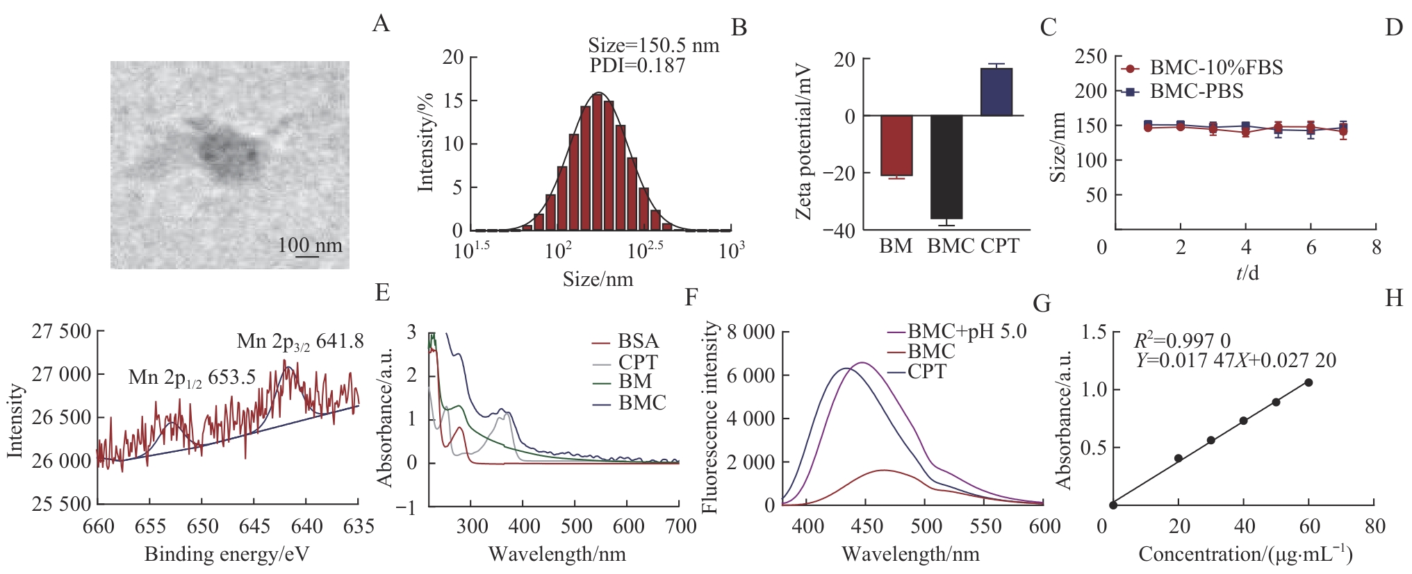
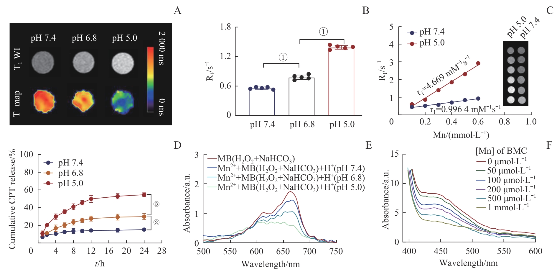
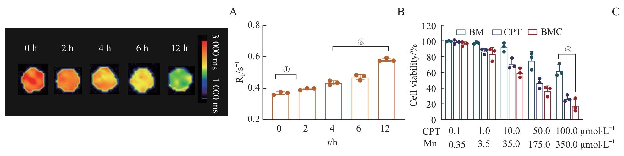
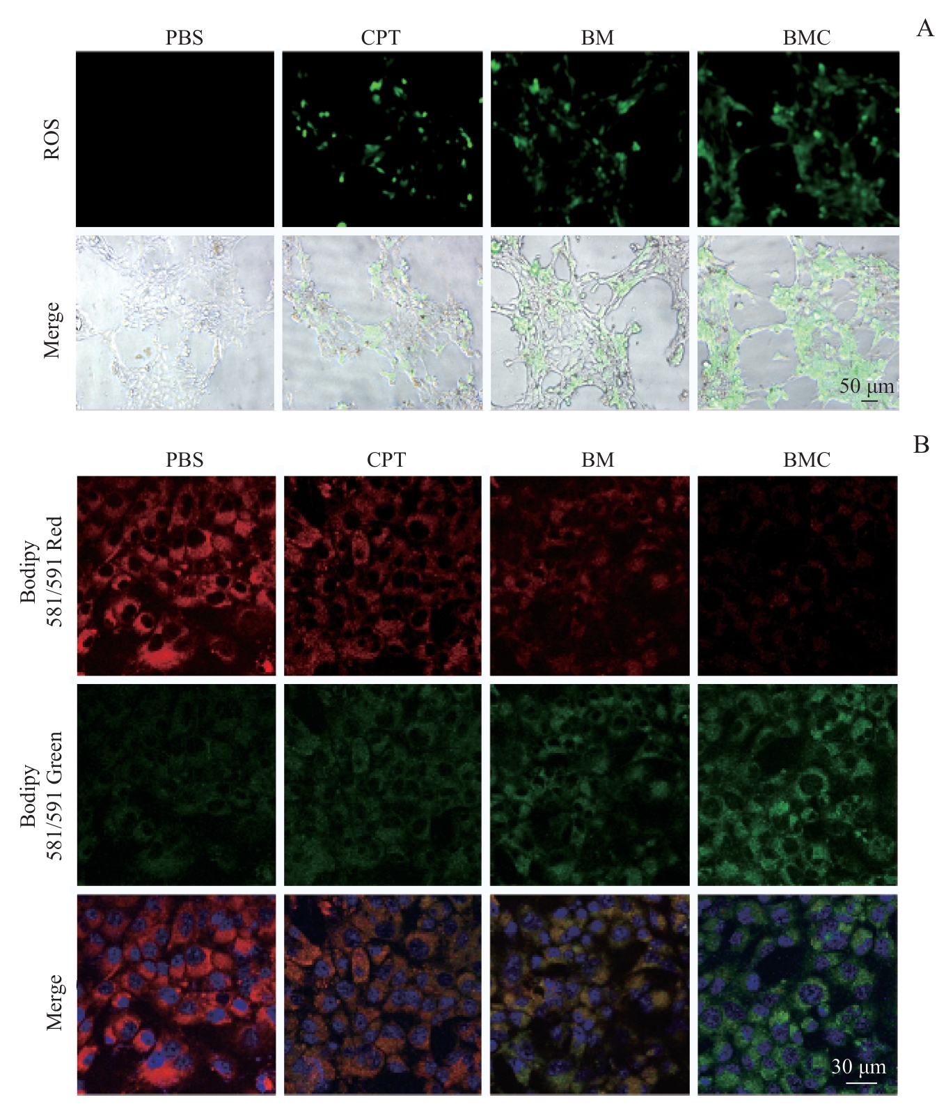
| [1] |
DE ROSE F, MEDURI B, DE SANTIS M C, et al. Rethinking breast cancer follow-up based on individual risk and recurrence management[J]. Cancer Treat Rev, 2022, 109: 102434.
|
| [2] |
LIU Y C, SUN Q Y, GUO J W, et al. Dual ferroptosis induction in N2-TANs and TNBC cells via FTH1 targeting: a therapeutic strategy for triple-negative breast cancer[J]. Cell Rep Med, 2025, 6(1): 101915.
|
| [3] |
JIN X, TANG J, QIU X, et al. Ferroptosis: emerging mechanisms, biological function, and therapeutic potential in cancer and inflammation[J]. Cell Death Discov, 2024, 10(1): 45.
|
| [4] |
HE H, DU L, XUE H, et al. Triple tumor microenvironment-responsive ferroptosis pathways induced by manganese-based imageable nanoenzymes for enhanced breast cancer theranostics[J]. Small Methods, 2023, 7(7): e2300230.
|
| [5] |
ZENG L, DING S, CAO Y, et al. A MOF-based potent ferroptosis inducer for enhanced radiotherapy of triple negative breast cancer[J]. ACS Nano, 2023, 17(14): 13195-13210.
|
| [6] |
DAI X, ZHU Y Q, SU M, et al. Rigid shell decorated nanodevice with Fe/H2O2 supply and glutathione depletion capabilities for potentiated ferroptosis and synergized immunotherapy[J]. Adv Funct Materials, 2023, 33(27):2215022.
|
| [7] |
LIU Y, PI F, HE L, et al. Oxygen vacancy-rich manganese nanoflowers as ferroptosis inducers for tumor radiotherapy[J]. Small, 2024, 20(32): e2310118.
|
| [8] |
ZHOU H, LU X, DU C, et al. Cycloacceleration of reactive oxygen species generation based on exceedingly small magnetic iron oxide nanoparticles for tumor ferroptosis therapy[J]. Small, 2022, 18(35): e2202705.
|
| [9] |
PAN S Y, SUN Z W, ZHAO B, et al. Therapeutic application of manganese-based nanosystems in cancer radiotherapy[J]. Biomaterials, 2023, 302: 122321.
|
| [10] |
LIN L S, SONG J, SONG L, et al. Simultaneous Fenton-like ion delivery and glutathione depletion by MnO2-based nanoagent to enhance chemodynamic therapy[J]. Angew Chem Int Ed, 2018, 57(18): 4902-4906.
|
| [11] |
CHENG J, ZHU Y, XING X, et al. Manganese-deposited iron oxide promotes tumor-responsive ferroptosis that synergizes the apoptosis of cisplatin[J]. Theranostics, 2021, 11(11): 5418-5429.
|
| [12] |
ZHU C D, MA Q, GONG L D, et al. Manganese-based multifunctional nanoplatform for dual-modal imaging and synergistic therapy of breast cancer[J]. Acta Biomater, 2022, 141: 429-439.
|
| [13] |
GAO Y, OUYANG Z, SHEN S, et al. Manganese dioxide-entrapping dendrimers co-deliver protein and nucleotide for magnetic resonance imaging-guided chemodynamic/starvation/immune therapy of tumors[J]. ACS Nano, 2023, 17(23): 23889-23902.
|
| [14] |
YOU J, LIU S K, LIANG J R, et al. Adaptive dual-responsive nanocapsules for precision ferroptosis-driven and chemotherapy-enhanced tumor ablation[J]. Med Mat, 2024, 1(2): 104-117.
|
| [15] |
朱仪, 邓佳丽, 王静怡, 等. Fe-MOF纳米探针在乳腺癌光动力增敏铁死亡及MR激活成像中实验研究[J]. 现代生物医学进展, 2024, 24(9): 1601-1607.
|
|
ZHU Y, DENG J L, WANG J Y, et al. Experimental study of Fe-MOF nanoprobe in photodynamically sensitized ferroptosis and MR activation imaging of breast cancer[J]. Progresss in Modern Biomedicine, 2024, 24(9): 1601-1607.
|
| [16] |
CHEN M Y, TONG X H, SUN Y T, et al. A ferroptosis amplifier based on triple-enhanced lipid peroxides accumulation strategy for effective pancreatic cancer therapy[J]. Biomaterials, 2024, 309: 122574.
|
| [17] |
LIU Q L, ZHAO Y L, ZHOU H G, et al. Ferroptosis: challenges and opportunities for nanomaterials in cancer therapy open access[J]. Regen Biomater, 2023, 10: rbad004.
|
| [18] |
DENG X, LIU T Z, ZHU Y T, et al. Ca & Mn dual-ion hybrid nanostimulator boosting anti-tumor immunity via ferroptosis and innate immunity awakening[J]. Bioact Mater, 2024, 33: 483-496.
|
| [19] |
ZHU Y, DENG J L, LU H W, et al. Reverse magnetic resonance tuning nanoplatform with heightened sensitivity for non-invasively multiscale visualizing ferroptosis-based tumor sensitization therapy[J]. Biomaterials, 2025, 315: 122935.
|
| [20] |
ZHENG H, HUANG L, AN G, et al. A nanoreactor based on metal-organic frameworks with triple synergistic therapy for hepatocellular carcinoma[J]. Adv Healthc Mater, 2024, 13(28): e2401743.
|
| [21] |
SUN Z, WANG Z, WANG T, et al. Biodegradable MnO-based nanoparticles with engineering surface for tumor therapy: simultaneous Fenton-like ion delivery and immune activation[J]. ACS Nano, 2022, 16(8): 11862-11875.
|
| [22] |
GIMENEZ C, ALPERIN M, DE VITA R. The effect of menopause on vaginal tissue mechanics: a brief review[J]. J Biomech Eng, 2024, 146(6): 060903.
|
| [23] |
许俊, 赵茜茜, 许乙凯. 高浓度磁共振对比剂钆布醇的理化性质及临床应用[J]. 放射学实践, 2016, 31(7): 666-669.
|
|
XU J, ZHAO Q Q, XU Y K. Physicochemical properties and clinical applications of the high-concentration MRI contrast agent gadobutrol [J]. Radiologic Practice, 2016, 31(7): 666-669.
|
| [24] |
WANG P, YANG J, ZHOU B, et al. Antifouling manganese oxide nanoparticles: synthesis, characterization, and applications for enhanced MR imaging of tumors[J]. ACS Appl Mater Interfaces, 2017, 9(1): 47-53.
|
 )
)