
上海交通大学学报(医学版) ›› 2026, Vol. 46 ›› Issue (8): 1053-1066.doi: 10.3969/j.issn.1674-8115.2026.08.006
• 论著 · 基础研究 • 上一篇
吴康卉1, 吴烁2, 罗成3, 张元元3( ), 李光明1()
收稿日期:2026-03-24
接受日期:2026-05-11
出版日期:2026-08-06
发布日期:2026-08-06
通讯作者:
李光明,主任医师,博士;电子信箱:liguangming@xinhuamed.com.cn作者简介:第一联系人:李光明和张元元负责课题的设计与指导,吴康卉与吴烁完成了实验操作、数据分析和文章撰写,罗成和张元元负责实验指导和论文修改。所有作者均阅读并同意了最终稿件的提交。基金资助:
Wu Kanghui1, Wu Shuo2, Luo Cheng3, Zhang Yuanyuan3(), Li Guangming1()
Received:2026-03-24
Accepted:2026-05-11
Online:2026-08-06
Published:2026-08-06
Contact:
Li Guangming, E-mail: liguangming@xinhuamed.com.cnAbout author:First author contact:Li Guangming and Zhang Yuanyuan were responsible for the project design and supervision. Wu Kanghui and Wu Shuo performed the experiments, conducted the data analysis, and wrote the manuscript. Luo Cheng and Zhang Yuanyuan were responsible for experimental guidance and manuscript revision. All authors have read the last version of paper and consented to its submission.
Supported by:摘要:
目的·探讨组蛋白赖氨酸去甲基化酶4B(lysine demethylase 4B,KDM4B)在结直肠癌(colorectal cancer,CRC)抗肿瘤免疫中的作用及其分子机制。方法·采用免疫组织化学染色(immunohistochemistry,IHC)检测CRC组织芯片中肿瘤组织和癌旁组织的KDM4B表达水平。利用基因表达综合数据库(Gene Expression Omnibus,GEO)的转录组测序数据集,分析KDM4B mRNA表达量与CRC患者预后、肿瘤内CD8+ T细胞浸润程度的相关性,并对KDM4B高表达组和低表达组的CRC样本进行基因集富集分析(Gene Set Enrichment Analysis,GSEA),以探索KDM4B相关的生物学过程。在CRC细胞中敲低KDM4B或使用KDM4B抑制剂B3处理,通过实时荧光定量PCR(quantitative real-time PCR,qPCR)、免疫荧光染色(immunofluorescence,IF)和蛋白质印迹法(Western blotting)检测该处理后细胞中KDM4B相关生物学过程。采用小鼠皮下成瘤实验评估敲低Kdm4b或B3处理在体内对CT26肿瘤增殖能力、肿瘤内CD8+ T细胞浸润水平和细胞毒性的影响,并且比较B3治疗、程序性死亡受体-1(programmed cell death protein-1,PD-1)抗体治疗及B3+PD-1抗体联合治疗对小鼠CT26皮下瘤的抗肿瘤作用。结果·IHC结果显示,CRC组织中的KDM4B表达明显高于癌旁组织(P<0.001)。GEO数据集分析的结果显示,KDM4B低表达组的CRC患者表现为更长的生存期以及更高的瘤内CD8+ T细胞浸润度(均P<0.05)。GSEA结果显示,KDM4B低表达与Ⅰ型干扰素反应、Th1型细胞毒性反应显著相关。qPCR、IF和蛋白质印迹法的结果显示,敲低KDM4B或B3处理在CRC细胞中能够促进DNA双链断裂、环鸟苷酸-腺苷酸合酶-干扰素基因刺激因子(cyclic GMP-AMP synthase-stimulator of interferon genes,cGAS-STING)信号通路激活、Ⅰ型干扰素和Th1型趋化因子表达增多(均P<0.05)。体内实验显示,敲低Kdm4b或B3处理能够限制CT26肿瘤的生长、促进瘤内CD8+ T细胞的浸润和颗粒酶B的表达(均P<0.05);相较于B3或PD-1抗体单药治疗,B3+PD-1抗体联合治疗对小鼠CT26皮下瘤的抗肿瘤作用更强(均P<0.05)。结论·敲低KDM4B或B3处理可增强CRC细胞的Ⅰ型干扰素和Th1型趋化因子表达,并在体内实验中促进CD8+ T细胞抗肿瘤免疫以提高CRC对PD-1抗体的敏感性。
中图分类号:
吴康卉, 吴烁, 罗成, 张元元, 李光明. KDM4B在结直肠癌中调控抗肿瘤免疫的机制研究[J]. 上海交通大学学报(医学版), 2026, 46(8): 1053-1066.
Wu Kanghui, Wu Shuo, Luo Cheng, Zhang Yuanyuan, Li Guangming. Mechanistic study of KDM4B in regulating anti-tumor immunity in colorectal cancer[J]. Journal of Shanghai Jiao Tong University (Medical Science), 2026,(8): 1053-1066.
| shRNA | Target sequence (5'→3') |
|---|---|
| shControl | TTCTCCGAACGTGTCACGT |
| shKdm4b#1 | GCCAGATCGTCATCACCAAGA |
| shKdm4b#2 | GCTGTAAGATCATGACTTTCC |
| shKDM4B#1 | GCGGCAGACGTATGATGACAT |
| shKDM4B#2 | GCCGGAAGGACATGGTCAAGA |
表1 shRNA目的序列
Tab 1 Target sequences of shRNA
| shRNA | Target sequence (5'→3') |
|---|---|
| shControl | TTCTCCGAACGTGTCACGT |
| shKdm4b#1 | GCCAGATCGTCATCACCAAGA |
| shKdm4b#2 | GCTGTAAGATCATGACTTTCC |
| shKDM4B#1 | GCGGCAGACGTATGATGACAT |
| shKDM4B#2 | GCCGGAAGGACATGGTCAAGA |
| Gene | Forward primer (5'→3') | Reverse primer (5'→3') |
|---|---|---|
| Ifn-α | TGCCCAGCAGATCAAGAAGG | TCAGGGGAAATTCCTGCACC |
| Ifn-β | AGCTCCAAGAAAGGACGAACA | GCCCTGTAGGTGAGGTTGAT |
| Cxcl9 | TGGAGTTCGAGGAACCCTAGT | AGGCAGGTTTGATCTCCGTT |
| Cxcl10 | CCAAGTGCTGCCGTCATTTTC | GGCTCGCAGGGATGATTTCAA |
| Cxcl11 | CCACGCTACCTTCTGTGGTT | ATGTTCGTGTGCCTCGTGAT |
| Actb | GCAAGTGCTTCTAGGCGGAC | AAGAAAGGGTGTAAAACGCAGC |
表 2 鼠源基因的qPCR引物序列
Tab 2 Primer sequences for qPCR of mouse genes
| Gene | Forward primer (5'→3') | Reverse primer (5'→3') |
|---|---|---|
| Ifn-α | TGCCCAGCAGATCAAGAAGG | TCAGGGGAAATTCCTGCACC |
| Ifn-β | AGCTCCAAGAAAGGACGAACA | GCCCTGTAGGTGAGGTTGAT |
| Cxcl9 | TGGAGTTCGAGGAACCCTAGT | AGGCAGGTTTGATCTCCGTT |
| Cxcl10 | CCAAGTGCTGCCGTCATTTTC | GGCTCGCAGGGATGATTTCAA |
| Cxcl11 | CCACGCTACCTTCTGTGGTT | ATGTTCGTGTGCCTCGTGAT |
| Actb | GCAAGTGCTTCTAGGCGGAC | AAGAAAGGGTGTAAAACGCAGC |
| Gene | Forward primer (5'→3') | Reverse primer (5'→3') |
|---|---|---|
| IFN-α | TCGTATGCCAGCTCACCTTT | CAGTCAGCATGGTCCTCTGT |
| IFN-β | GGCACAACAGGTAGTAGGCG | GTGGAGAAGCACAACAGGAGA |
| CXCL9 | GTGGTGTTCTTTTCCTCTTGGG | ACAGCGACCCTTTCTCACTAC |
| CXCL10 | GCTTCCAAGGATGGACCACA | GCAGGGTCAGAACATCCACT |
| CXCL11 | CTGCCCAAAGGAGTCCAACA | TGTCTCCACCGTAACCACAG |
| β-actin | GAGAAAATCTGGCACCACACC | ATACCCCTCGTAGATGGGCAC |
表 3 人源基因的qPCR引物序列
Tab 3 Primer sequences for qPCR of human genes
| Gene | Forward primer (5'→3') | Reverse primer (5'→3') |
|---|---|---|
| IFN-α | TCGTATGCCAGCTCACCTTT | CAGTCAGCATGGTCCTCTGT |
| IFN-β | GGCACAACAGGTAGTAGGCG | GTGGAGAAGCACAACAGGAGA |
| CXCL9 | GTGGTGTTCTTTTCCTCTTGGG | ACAGCGACCCTTTCTCACTAC |
| CXCL10 | GCTTCCAAGGATGGACCACA | GCAGGGTCAGAACATCCACT |
| CXCL11 | CTGCCCAAAGGAGTCCAACA | TGTCTCCACCGTAACCACAG |
| β-actin | GAGAAAATCTGGCACCACACC | ATACCCCTCGTAGATGGGCAC |
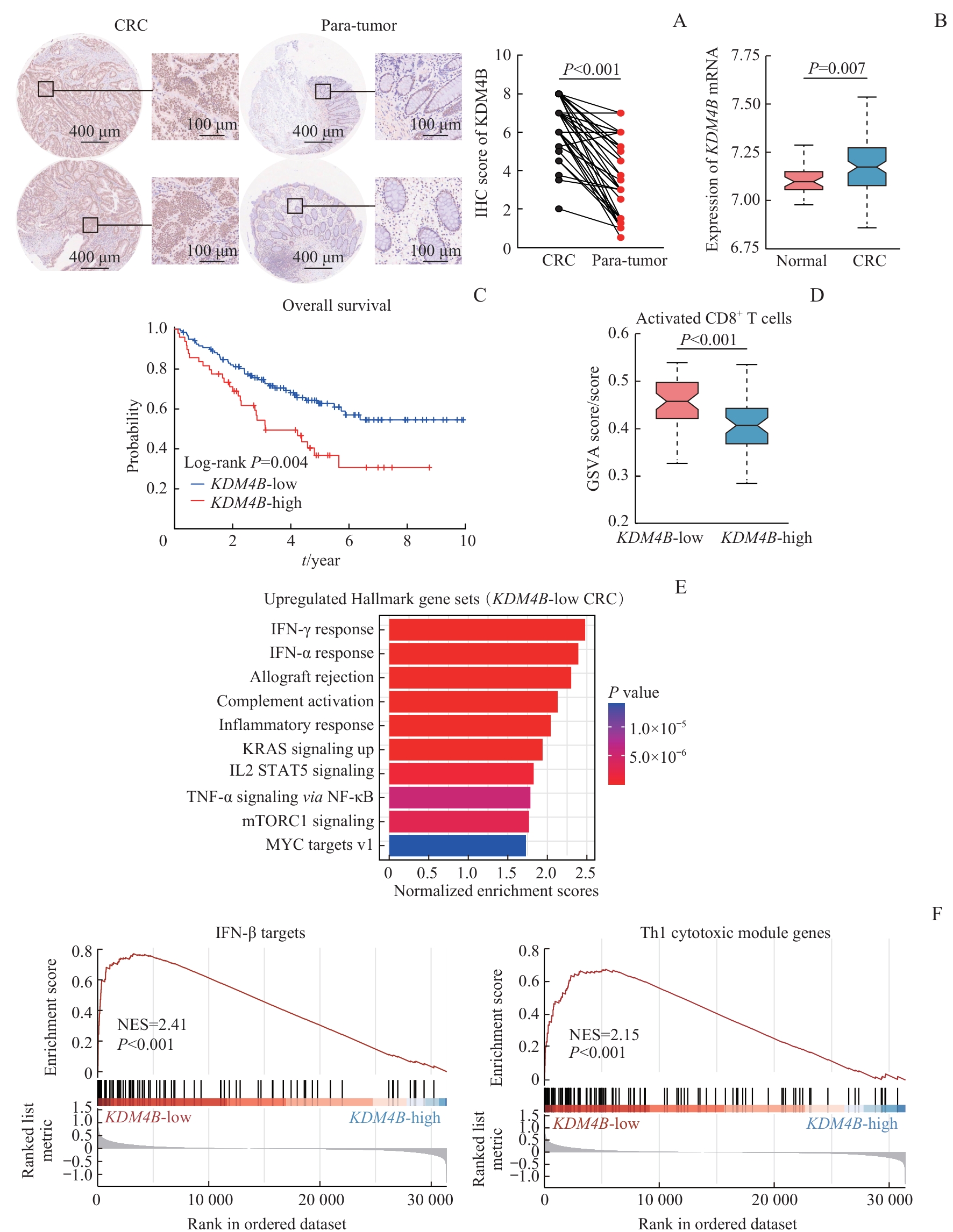
图1 KDM4B在CRC组织中的表达及其与患者预后、CD8+ T细胞浸润的关系Note: A. KDM4B expression in CRC tissues and para-tumor tissues detected by IHC staining (left: ×5; right: ×20). B. Expression of KDM4B mRNA in CRC and normal specimens from the GSE83889 dataset. C. Overall survival curves of CRC patients stratified by high versus low KDM4B expression in the GSE17536 dataset. D. GSVA scores of activated CD8+ T cells between KDM4B-high and KDM4B-low expression specimens in the GSE83889 dataset. E. The ten most significantly upregulated Hallmark gene sets in CRC samples with low KDM4B expression compared with those with high KDM4B expression identified by GSEA. F. Enrichment analysis of IFN-β targets and Th1 cytotoxic module genes sets in the KDM4B-low expression group versus the KDM4B-high expression group by GSEA. KRAS—kirsten rat sarcoma viral oncogene homolog; IL2—interleukin-2; STAT5—signal transducer and activator of transcription 5; TNF-α—tumour necrosis factor-α; mTORC1—mechanistic target of rapamycin complex 1; MYC—myelocytomatosis oncogene gene; NF-κB—nuclear factor kappa-B; NES—normalized enrichment score.
Fig 1 Expression of KDM4B in CRC tissue and its correlations with patient prognosis and CD8+ T cell infiltration
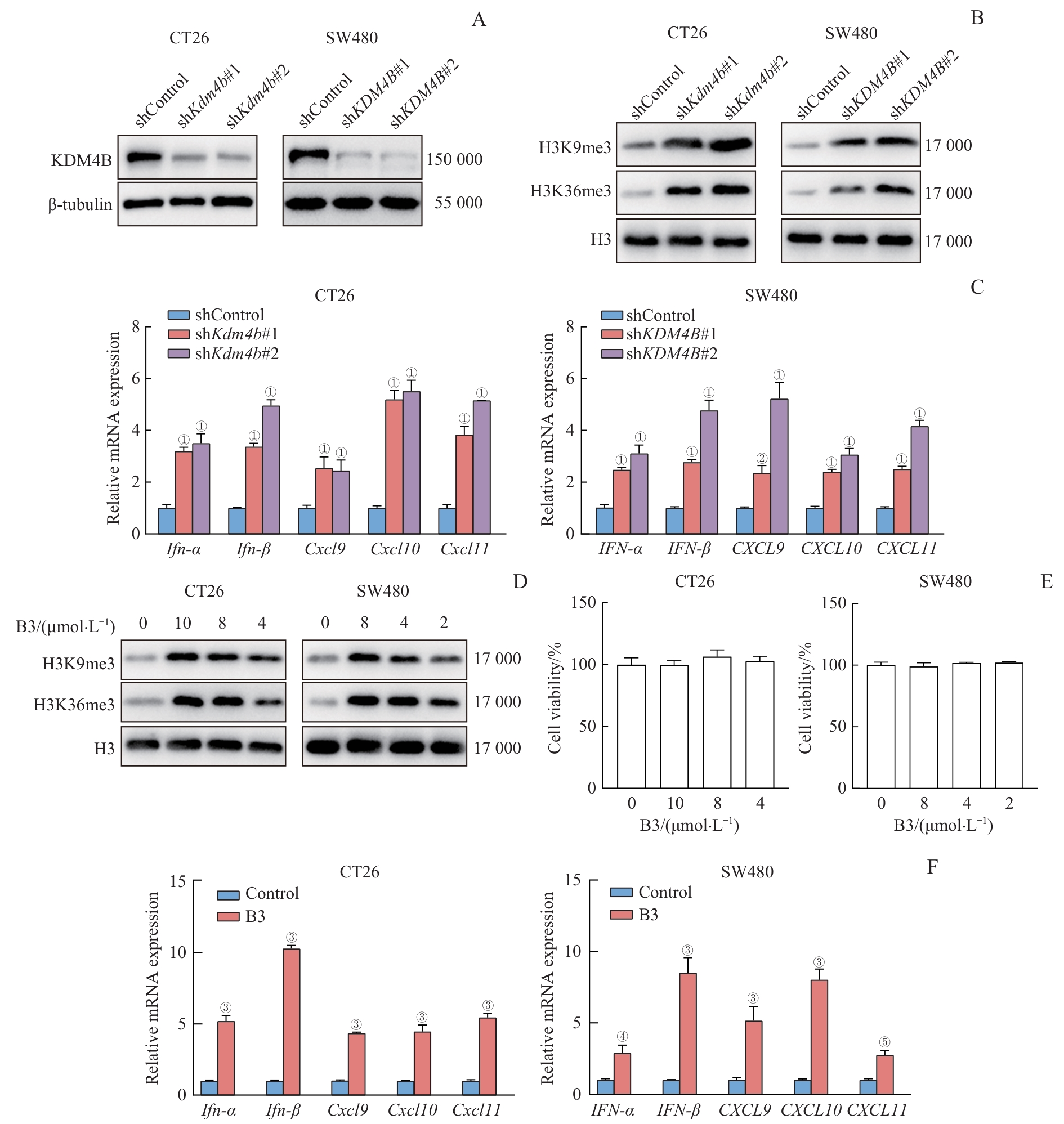
图2 抑制KDM4B对Ⅰ型干扰素和趋化因子表达水平的影响Note: A. Detection of the efficacy of KDM4B knockdown in CT26 and SW480 cells by Western blotting. B. Detection of H3K9me3 and H3K36me3 expression in CT26 or SW480 cells after KDM4B knockdown by Western blotting. C. Relative mRNA expression levels of the interferons IFN-α/IFN-β and chemokines CXCL9/10/11 in CT26 or SW480 cells following KDM4B knockdown by qPCR. D. Detection of H3K9me3 and H3K36me3 expression in CT26 or SW480 cells afterB3 treatment by Western blotting. E. Cell viability of CT26 or SW480 cells treated with different concentrations of B3.F. Relative mRNA expression levels of the interferons IFN-α/IFN-β and chemokines CXCL9/10/11 in CT26 or SW480 cells followingB3 treatment by qPCR. ①P<0.001, ②P=0.001, compared with the shControl group; ③P<0.001, ④P=0.003, ⑤P=0.006, compared with the Control group.
Fig 2 Effects of KDM4B inhibition on the expression levels of type Ⅰinterferons and Th1 type chemokines
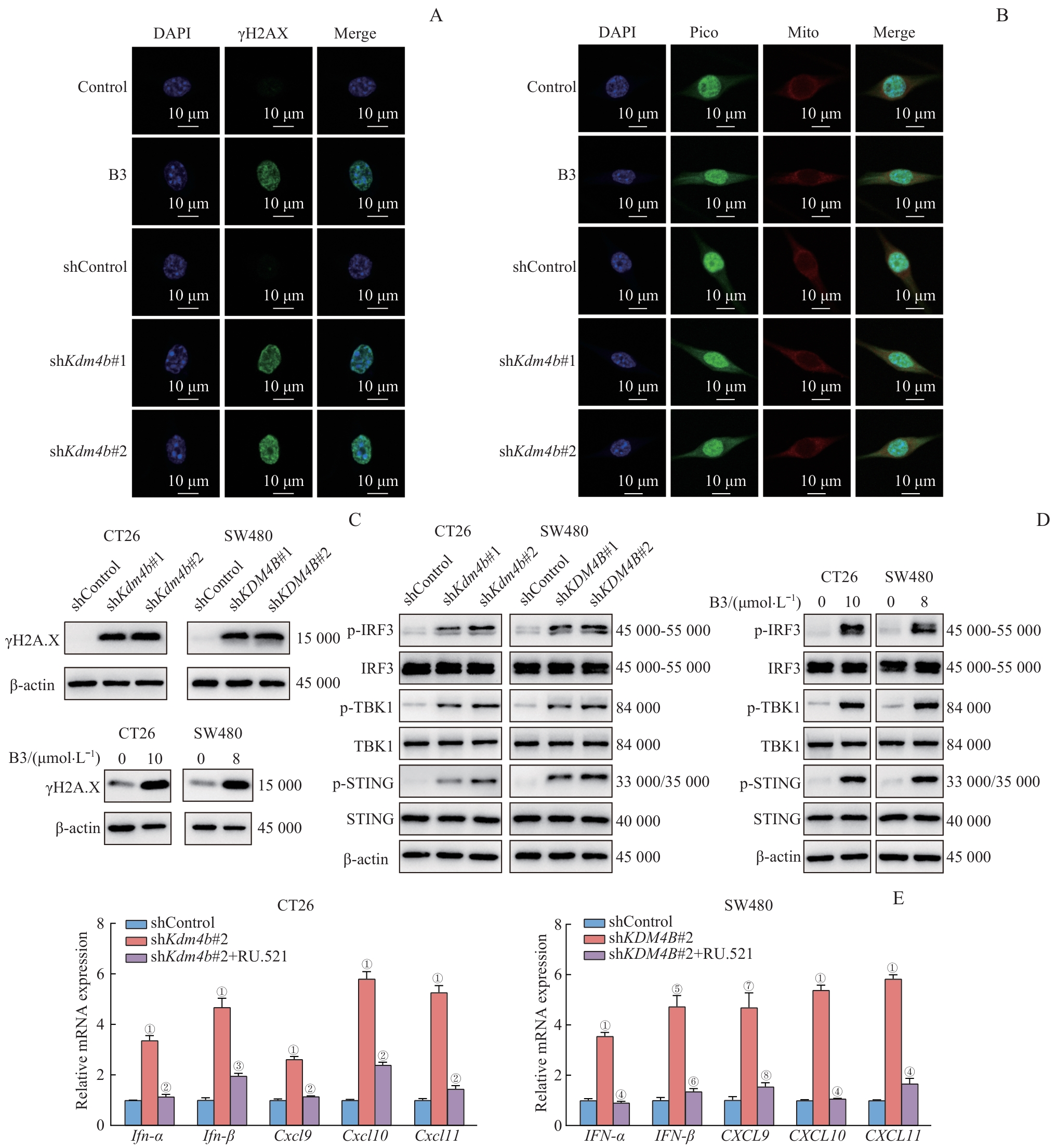
图3 敲低 KDM4B 或B3处理对DNA损伤和cGAS-STING信号轴的影响Note: A. IF staining of γH2A.X (green) in CT26 cells subjected to Kdm4b knockdown or B3 treatment (×63). B. Fluorescence staining of dsDNA (green) and mitochondria (red) in CT26 cells subjected to Kdm4b knockdown or B3 treatment (×63). C. Detection of γH2A.X protein expression in CT26 or SW480 cells after KDM4B knockdown or B3 treatment by Western blotting. D. Detection of the protein expression levels of cGAS-STING axis components in CT26 or SW480 cells following KDM4B knockdown or B3 treatment by Western blotting. F. Relative mRNA expression levels of IFN-α/IFN-β and chemokines CXCL9/10/11 in KDM4B-knockdown CT26 or SW480 cells treated with RU.521. ①P<0.001, ⑤P=0.001, ⑦P=0.004, compared with the shControl group; ②P<0.001, ③P=0.002, compared with the shKdm4b#2 group; ④P<0.001, ⑥P=0.002, ⑧P=0.006, compared with the shKDM4B#2 group.
Fig 3 Effects of KDM4B knockdown or B3 treatment on DNA damage and the cGAS-STING signaling axis
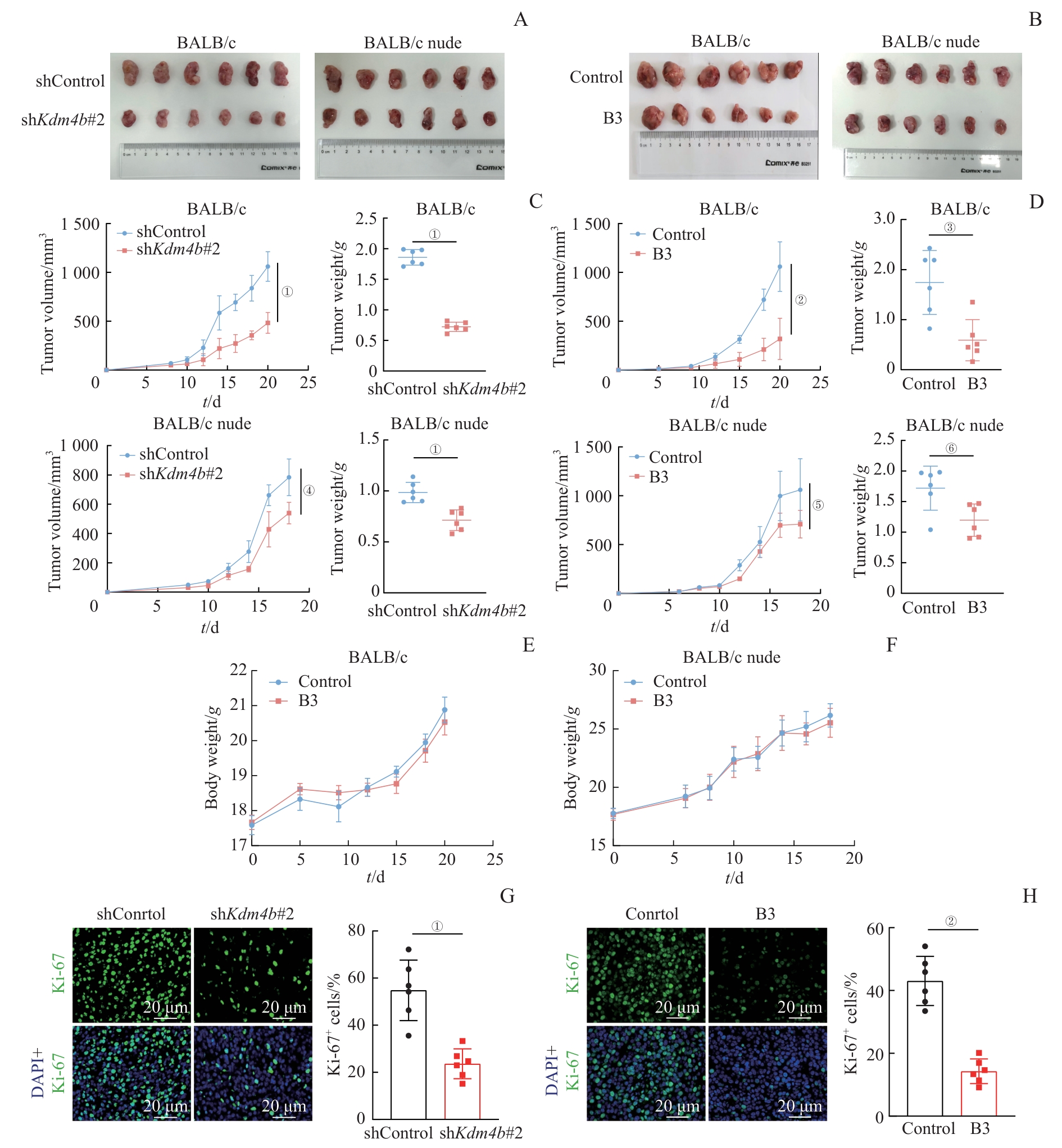
图4 敲低 Kdm4b 或B3处理后CT26细胞体内成瘤和增殖能力的变化Note: A/B. CT26 tumors derived from BALB/c and BALB/c nude mice following Kdm4b knockdown (A) or B3 treatment (B). C/D. Tumor volume and weight of BALB/c and BALB/c nude mice following Kdm4b knockdown (C) or B3 treatment (D). E/F. Body weight of BALB/c mice (E) or BALB/c nude mice (F) implanted with CT26 cells after intratumoral injection of vehicle or B3. G/H. IF staining of Ki-67 in CT26 tumor tissues after Kdm4b knockdown (G) or B3 treatment (H) in BALB/c mice (×20). ①P<0.001, ④P=0.002, compared with the shControl group; ②P<0.001, ③P=0.003, ⑤P=0.033, ⑥P=0.017, compared with the Control group.
Fig 4 Tumorigenic and proliferative capacities of CT26 cells in vivo following Kdm4b knockdown or B3 treatment
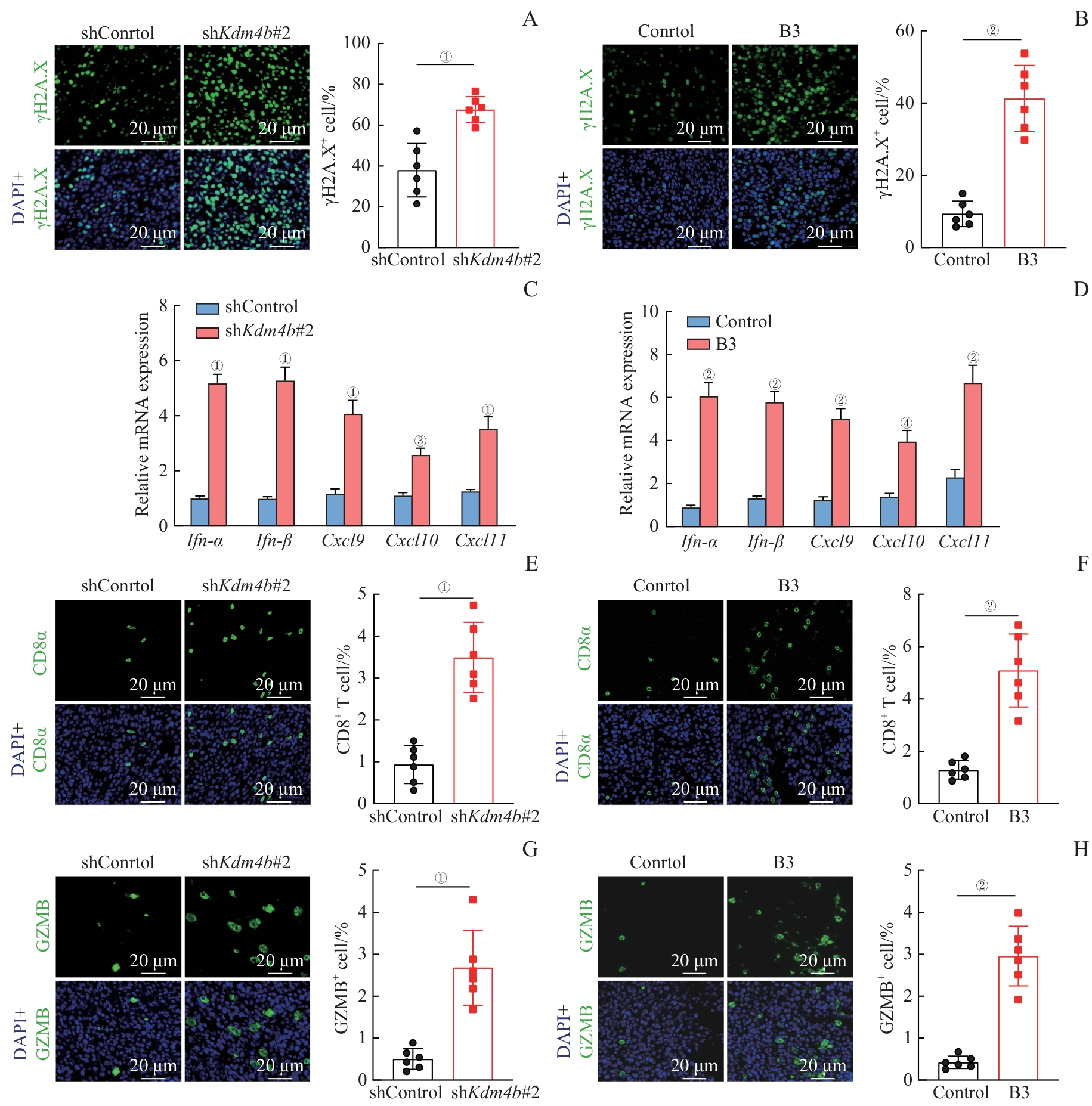
图5 体内研究中敲低 Kdm4b 或B3处理对DNA损伤、CD8+ T细胞浸润和GZMB表达的影响Note: A/B. Detection of γH2A.X expression in tumor tissues from BALB/c mice with Kdm4b knockdown (A) or B3 treatment (B) by IF staining (×20). C/D. Relative mRNA expression levels of type Ⅰ interferons Ifn-α/Ifn-β and Th1 type chemokines Cxcl9/10/11 in tumor tissues from BALB/c mice with Kdm4b knockdown (C) or B3 treatment (D) detected by qPCR. E/F. Detection of CD8⁺ T cells infiltration in CT26 tumor tissues after Kdm4b knockdown (E) or B3 treatment (F) in BALB/c mice by IF staining (×20). G/H. Detection of GZMB expression in CT26 tumor tissues after Kdm4b knockdown (G) or B3 treatment (H) in BALB/c mice by IF staining (×20). ①P<0.001, ③P=0.006, compared with the shControl group; ②P<0.001, ④P=0.001, compared with the Control group.
Fig 5 Effects of Kdm4b knockdown or B3 treatment on DNA damage, CD8⁺ T cells infiltration, and GZMB expression in vivo
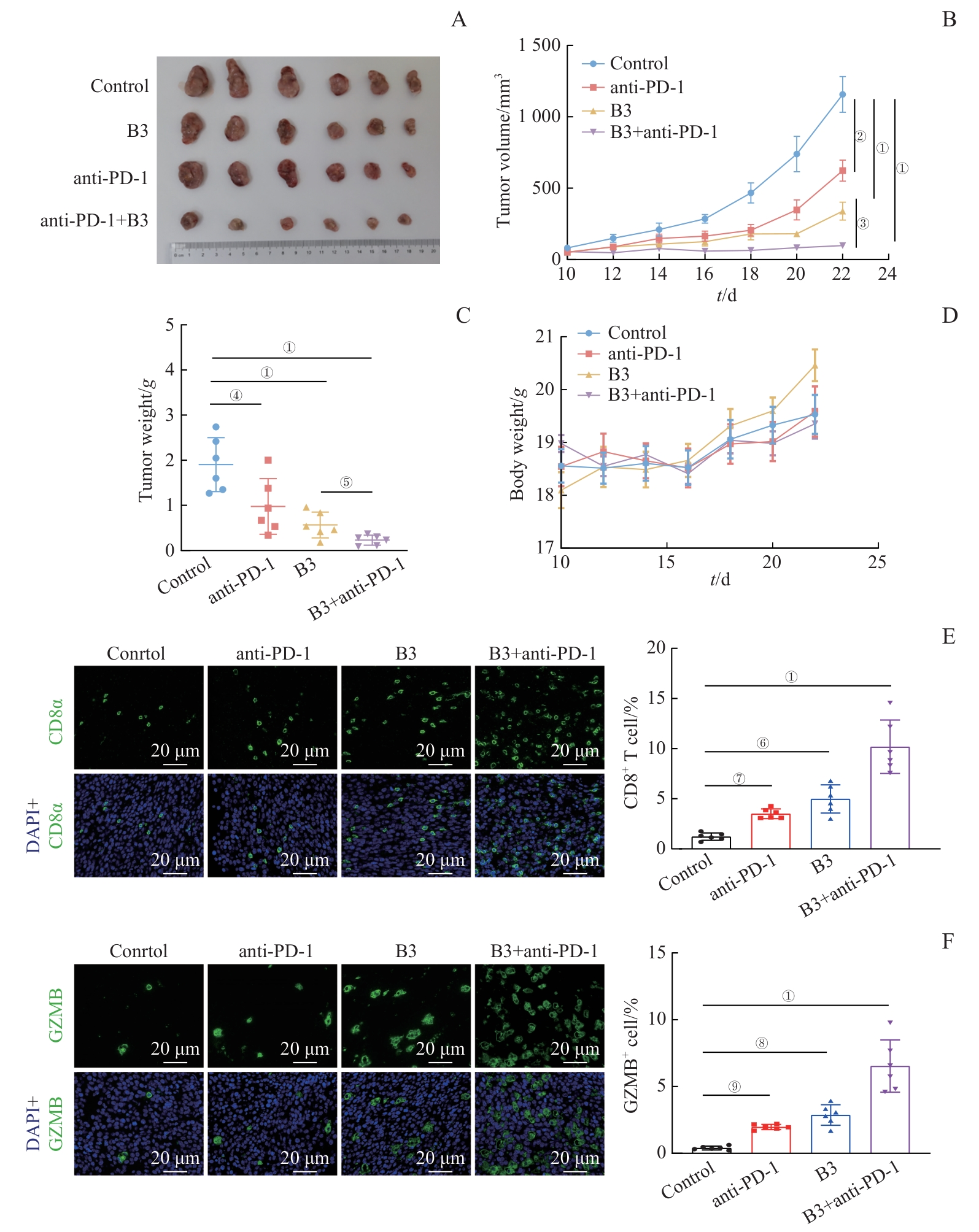
图6 体内研究中B3联合PD-1抗体治疗对CT26肿瘤生长、肿瘤内CD8+ T细胞浸润和GZMB表达的影响Note: A. CT26 tumors derived from BALB/c mice among the four groups (Control, anti-PD-1, B3, and B3+anti-PD-1). B/C. Tumor volume (B) and weight (C) of BALB/c mice subcutaneously implanted with CT26 cells in each group. D.Body weight of BALB/c mice subcutaneously implanted with CT26 cells in each group. E/F. IF staining of CD8⁺ T cells (E) and GZMB+ cells (F) in CT26 tumor tissues among the four groups (×20). ①P<0.001, ②P=0.004, ④P=0.006, ⑥P=0.001, ⑦P=0.047, ⑧P=0.002, ⑨P=0.046, compared with the Control group; ③P=0.004, ⑤P=0.023, B3 group vs B3+anti-PD-1 group.
Fig 6 Effects of B3 combined with anti-PD-1 antibody therapy on CT26 tumor growth, intratumoral CD8⁺ T cells infiltration, and GZMB expression in vivo
| [1] | Ahuja S, Zaheer S. The evolution of cancer immunotherapy: a comprehensive review of its history and current perspectives[J]. Korean J Clin Oncol, 2024, 20(2): 51-73. |
| [2] | Rui R, Zhou L Q, He S M. Cancer immunotherapies: advances and bottlenecks[J]. Front Immunol, 2023, 14: 1212476. |
| [3] | Yamaguchi H, Hsu J M, Sun L L, et al. Advances and prospects of biomarkers for immune checkpoint inhibitors[J]. Cell Rep Med, 2024, 5(7): 101621. |
| [4] | Salva de Torres C, Baraibar I, Saoudi González N, et al. Current and emerging treatment paradigms in colorectal cancer: integrating hallmarks of cancer[J]. Int J Mol Sci, 2024, 25(13): 6967. |
| [5] | Abedizadeh R, Majidi F, Khorasani H R, et al. Colorectal cancer: a comprehensive review of carcinogenesis, diagnosis, and novel strategies for classified treatments[J]. Cancer Metastasis Rev, 2024, 43(2): 729-753. |
| [6] | Siegel R L, Wagle N S, Cercek A, et al. Colorectal cancer statistics, 2023[J]. CA Cancer J Clin, 2023, 73(3): 233-254. |
| [7] | Shan J Q, Han D, Shen C Y, et al. Mechanism and strategies of immunotherapy resistance in colorectal cancer[J]. Front Immunol, 2022, 13: 1016646. |
| [8] | Yan S Y, Wang W T, Feng Z Q, et al. Immune checkpoint inhibitors in colorectal cancer: limitation and challenges[J]. Front Immunol, 2024, 15: 1403533. |
| [9] | Yang J, Xu J, Wang W, et al. Epigenetic regulation in the tumor microenvironment: molecular mechanisms and therapeutic targets[J]. Signal Transduct Target Ther, 2023, 8(1): 210. |
| [10] | Cao J, Yan Q. Cancer epigenetics, tumor immunity, and immunotherapy[J]. Trends Cancer, 2020, 6(7): 580-592. |
| [11] | Ni F J, Tang H T, Cheng S T, et al. KDM4B: a promising oncology therapeutic target[J]. Cancer Sci, 2024, 115(1): 8-16. |
| [12] | Wang Z Z, Cai H R, Zhao E H, et al. The diverse roles of histone demethylase KDM4B in normal and cancer development and progression[J]. Front Cell Dev Biol, 2021, 9: 790129. |
| [13] | Zhang N, Liu Y, Zhang X D, et al. Cetrorelix promotes cell apoptosis via the PI3K-AKT-FOXO1 pathway in epithelial ovarian cancer[J]. Front Oncol, 2025, 15: 1631576. |
| [14] | Qu F J, Wu S, Yu W W. Progress of immune checkpoint inhibitors therapy for pMMR/MSS metastatic colorectal cancer[J]. Onco-Targets Ther, 2024, 17: 1223-1253. |
| [15] | Fu L N, Chen L S, Yang J, et al. HIF-1α-induced histone demethylase JMJD2B contributes to the malignant phenotype of colorectal cancer cells via an epigenetic mechanism[J]. Carcinogenesis, 2012, 33(9): 1664-1673. |
| [16] | Chen Y X, Yu D N, Qian H, et al. CD8+ T cell-based cancer immunotherapy[J]. J Transl Med, 2024, 22(1): 394. |
| [17] | Wu B, Zhang B, Li B W, et al. Cold and hot tumors: from molecular mechanisms to targeted therapy[J]. Signal Transduct Target Ther, 2024, 9(1): 274. |
| [18] | Philip M, Schietinger A. CD8+ T cell differentiation and dysfunction in cancer[J]. Nat Rev Immunol, 2022, 22(4): 209-223. |
| [19] | Zannikou M, Fish E N, Platanias L C. Signaling by type I interferons in immune cells: disease consequences[J]. Cancers, 2024, 16(8): 1600. |
| [20] | Lim J, Kang I, La J, et al. Harnessing type I interferon-mediated immunity to target malignant brain tumors[J]. Front Immunol, 2023, 14: 1203929. |
| [21] | Tokunaga R, Zhang W, Naseem M, et al. CXCL9, CXCL10, CXCL11/CXCR3 axis for immune activation: a target for novel cancer therapy[J]. Cancer Treat Rev, 2018, 63: 40-47. |
| [22] | Jung H, Paust S. Chemokines in the tumor microenvironment: implications for lung cancer and immunotherapy[J]. Front Immunol, 2024, 15: 1443366. |
| [23] | Sulkowski P L, Oeck S, Dow J, et al. Oncometabolites suppress DNA repair by disrupting local chromatin signalling[J]. Nature, 2020, 582(7813): 586-591. |
| [24] | Zhang W C, Liu W, Jia L F, et al. Targeting KDM4A epigenetically activates tumor-cell-intrinsic immunity by inducing DNA replication stress[J]. Mol Cell, 2021, 81(10): 2148-2165.e9. |
| [25] | Yue B L, Gao W B, Lovell J F, et al. The cGAS-STING pathway in cancer immunity: dual roles, therapeutic strategies, and clinical challenges[J]. Essays Biochem, 2025, 69(2): EBC20253006. |
| [26] | Li J Y, Chen Y P, Li Y Q, et al. Chemotherapeutic and targeted agents can modulate the tumor microenvironment and increase the efficacy of immune checkpoint blockades[J]. Mol Cancer, 2021, 20(1): 27. |
| [1] | 翟元晖, 王莹莹, 王宇轩, 吴文萱, 朱楚娇, 张幼萍, 白雯会, 雷虎, 徐含章, 赵亚雪, 吴英理. 甲基巴多索隆作为新型泛素特异性蛋白酶48抑制剂抑制结直肠癌的作用及机制研究[J]. 上海交通大学学报(医学版), 2026, 46(8): 1026-1036. |
| [2] | 沈雨潇, 李晓光, 巴乾. SHP2抑制类药物的开发及在肿瘤治疗中的应用研究进展[J]. 上海交通大学学报(医学版), 2026, 46(6): 713-720. |
| [3] | 陈毓涵, 艾志宏. 子宫内膜癌的免疫微环境及免疫治疗研究进展[J]. 上海交通大学学报(医学版), 2026, 46(5): 656-664. |
| [4] | 江怡, 黄晨浩, 李祉良, 吴珺玮, 赵任, 张弢. 1例KRAS突变的结直肠癌患者术前接受化疗联合免疫治疗的效果报道[J]. 上海交通大学学报(医学版), 2025, 45(9): 1256-1260. |
| [5] | 孟靖, 谢玉婷, 左佳鑫, 熊屏. 纳米工程化T细胞体系的构建及其对口腔鳞状细胞癌的体外治疗研究[J]. 上海交通大学学报(医学版), 2025, 45(7): 866-873. |
| [6] | 陈子旋, 刘敏. 肾细胞癌免疫细胞治疗的研究进展[J]. 上海交通大学学报(医学版), 2025, 45(7): 916-925. |
| [7] | 陈佳莹, 褚以忞, 彭海霞. 结直肠癌无进展生存时间预测模型及影响因素研究[J]. 上海交通大学学报(医学版), 2025, 45(3): 324-334. |
| [8] | 张涤凡, 王明慧, 赵洁, 万江波, 黄方, 郝思国. B7基因修饰的白血病细胞外泌体的抗白血病效应[J]. 上海交通大学学报(医学版), 2025, 45(2): 129-137. |
| [9] | 孙瑞状, 乔坤朋, 李璞, 徐小莲, 孟俊. ABCA7在结直肠癌中的临床价值及转录调控机制[J]. 上海交通大学学报(医学版), 2025, 45(11): 1490-1501. |
| [10] | 梁乐斌, 陈慧芳, 赖淑静, 顾靓, 苏冰. 基于空间ATAC-seq技术的Apcmin/+小鼠结肠肿瘤表观特征分析[J]. 上海交通大学学报(医学版), 2025, 45(10): 1261-1270. |
| [11] | 唐珺倩, 李本尚. 儿童高危细胞遗传学B系急性淋巴细胞白血病治疗新进展[J]. 上海交通大学学报(医学版), 2025, 45(10): 1390-1399. |
| [12] | 钱立恒, 温凯玲, 廖颖娜, 李书鑫, 聂惠贞. 分选链接蛋白1抑制结直肠癌细胞增殖和迁移的作用和机制研究[J]. 上海交通大学学报(医学版), 2024, 44(9): 1124-1135. |
| [13] | 何蕊, 颜克鹏, 王静. 靶向髓源性抑制细胞的叶酸循环增强肿瘤免疫治疗效果研究[J]. 上海交通大学学报(医学版), 2024, 44(8): 1011-1022. |
| [14] | 胡飞, 蔡晓涵, 程睿, 季诗雨, 苗嘉欣, 朱晏, 范广建. 骨肉瘤免疫微环境及其免疫治疗临床转化研究进展[J]. 上海交通大学学报(医学版), 2024, 44(7): 814-821. |
| [15] | 冯昫皎, 刘健悦, 戚炀炀, 孙晶, 沈蕾. 结直肠癌中自然杀伤细胞表型及功能初探[J]. 上海交通大学学报(医学版), 2024, 44(6): 713-722. |
| 阅读次数 | ||||||
|
全文 |
|
|||||
|
摘要 |
|
|||||
